
- FASTA Sequence
- PDBx/mmCIF Format
- PDBx/mmCIF Format (gz)
- BinaryCIF Format (gz)
- Legacy PDB Format
- Legacy PDB Format (gz)
- PDBML/XML Format (gz)
- Structure Factors (CIF)
- Structure Factors (CIF - gz)
- Validation Full (PDF - gz)
- Validation (XML - gz)
- Validation (CIF - gz)
- Validation 2fo-fc coefficients (CIF - gz)
- Validation fo-fc coefficients (CIF - gz)
- Biological Assembly 1 (CIF - gz)
- Biological Assembly 1 (PDB - gz)
4O02 | pdb_00004o02
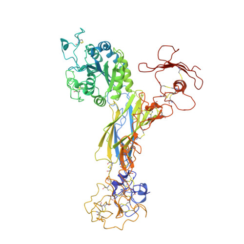
AlphaVBeta3 integrin in complex with monoclonal antibody FAB fragment.
- PDB DOI: https://doi.org/10.2210/pdb4O02/pdb
- Classification: PROTEIN BINDING
- Organism(s): Homo sapiens, Mus musculus
- Expression System: Spodoptera frugiperda
- Mutation(s): No
- Deposited: 2013-12-13 Released: 2014-04-02
- Deposition Author(s): Mahalingam, B., van Agthoven, J., Xiong, J., Arnaout, M.A.
Experimental Data Snapshot
- Method: X-RAY DIFFRACTION
- Resolution: 3.60 Å
- R-Value Free: 0.312 (Depositor), 0.314 (DCC)
- R-Value Work: 0.246 (Depositor), 0.254 (DCC)
- R-Value Observed: 0.250 (Depositor)
Literature
- 👁 Image
Download Mendeley
Atomic basis for the species-specific inhibition of alpha V integrins by monoclonal antibody 17E6 is revealed by the crystal structure of alpha V beta 3 ectodomain-17E6 Fab complex.
Mahalingam, B., Van Agthoven, J.F., Xiong, J.P., Alonso, J.L., Adair, B.D., Rui, X., Anand, S., Mehrbod, M., Mofrad, M.R., Burger, C., Goodman, S.L., Arnaout, M.A.(2014) J Biological Chem 289: 13801-13809
- PubMed: 24692540 Search on PubMedSearch on PubMed Central
- DOI: https://doi.org/10.1074/jbc.M113.546929
- Primary Citation Related Structures:
4O02 - PubMed Abstract:
The function-blocking, non-RGD-containing, and primate-specific mouse monoclonal antibody 17E6 binds the αV subfamily of integrins. 17E6 is currently in phase II clinical trials for treating cancer. To elucidate the structural basis of recognition and the molecular mechanism of inhibition, we crystallized αVβ3 ectodomain in complex with the Fab fragment of 17E6. Protein crystals grew in presence of the activating cation Mn(2+). The integrin in the complex and in solution assumed the genuflected conformation. 17E6 Fab bound exclusively to the Propeller domain of the αV subunit. At the core of αV-Fab interface were interactions involving Propeller residues Lys-203 and Gln-145, with the latter accounting for primate specificity. The Propeller residue Asp-150, which normally coordinates Arg of the ligand Arg-Gly-Asp motif, formed contacts with Arg-54 of the Fab that were expected to reduce soluble FN10 binding to cellular αVβ3 complexed with 17E6. This was confirmed in direct binding studies, suggesting that 17E6 is an allosteric inhibitor of αV integrins.
- From the Structural Biology Program and.
- the Leukocyte Biology and Inflammation Program, Departments of Medicine and Developmental & Regenerative Biology, Massachusetts General Hospital and Harvard Medical School, Charlestown, Massachusetts 02129.
- the Departments of Bioengineering and Mechanical Engineering, University of California, Berkeley, California 94720.
- Merck KGaA and Discovery Technologies, Molecular Pharmacology, and.
- Merck KGaA and Therapeutic Innovation Platform, Oncology, Darmstadt 64271, Germany.
- From the Structural Biology Program and the Leukocyte Biology and Inflammation Program, Departments of Medicine and Developmental & Regenerative Biology, Massachusetts General Hospital and Harvard Medical School, Charlestown, Massachusetts 02129, aarnaout1@mgh.harvard.edu.
Organizational Affiliation:
Explore in 3D: Structure | Sequence Annotations | Electron Density | Validation Report | Ligand Interaction (NAG)
Biological Assembly 1
Explore in 3D: Structure | Sequence Annotations | Electron Density | Validation Report | Ligand Interaction (NAG)
Global Symmetry: Asymmetric - C1
Global Stoichiometry: Hetero 4-mer - A1B1C1D1
Pseudo Symmetry: Asymmetric - C1
Pseudo Stoichiometry: Hetero 4-mer - A2B1C1
Find Similar Assemblies
Biological assembly 1 assigned by authors.
Macromolecule Content
- Total Structure Weight: 236.04 kDa
- Atom Count: 13,526
- Modeled Residue Count: 2,034
- Deposited Residue Count: 2,086
- Unique protein chains: 4
Macromolecules
Entity ID: 1 | |||||
|---|---|---|---|---|---|
| Molecule | Chains | Sequence Length | Organism | Details | Image |
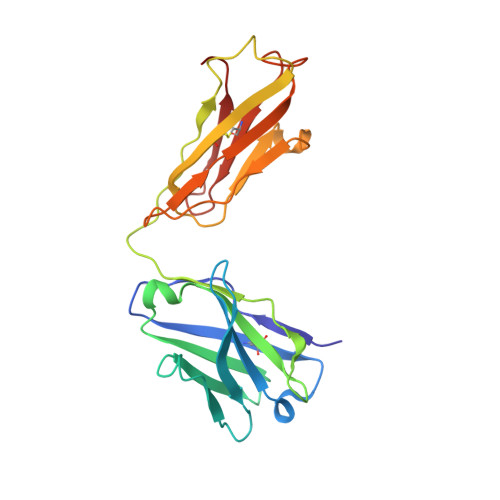
| Integrin alpha-V | 962 | Homo sapiens | Mutation(s): 0 Gene Names: ITGAV, MSK8, VNRA | 👁 Image | |
UniProt & NIH Common Fund Data Resources | |||||
PHAROS: P06756 GTEx: ENSG00000138448 | |||||
Entity Groups | |||||
| Sequence Clusters | 30% Identity50% Identity70% Identity90% Identity95% Identity100% Identity | ||||
| UniProt Group | P06756 | ||||
Glycosylation | |||||
| Glycosylation Sites: 6 | Go to GlyGen: P06756-1 | ||||
Sequence AnnotationsExpand | |||||
Reference Sequence | |||||
Entity ID: 2 | |||||
|---|---|---|---|---|---|
| Molecule | Chains | Sequence Length | Organism | Details | Image |
| Integrin beta-3 | 692 | Homo sapiens | Mutation(s): 0 Gene Names: ITGB3, GP3A | 👁 Image | |
UniProt & NIH Common Fund Data Resources | |||||
PHAROS: P05106 GTEx: ENSG00000259207 | |||||
Entity Groups | |||||
| Sequence Clusters | 30% Identity50% Identity70% Identity90% Identity95% Identity100% Identity | ||||
| UniProt Group | P05106 | ||||
Glycosylation | |||||
| Glycosylation Sites: 4 | Go to GlyGen: P05106-1 | ||||
Sequence AnnotationsExpand | |||||
Reference Sequence | |||||
Entity ID: 3 | |||||
|---|---|---|---|---|---|
| Molecule | Chains | Sequence Length | Organism | Details | Image |
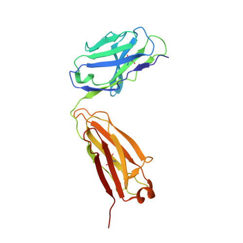
| 17E6 light chain | C [auth L] | 214 | Mus musculus | Mutation(s): 0 | 👁 Image |
Entity ID: 4 | |||||
|---|---|---|---|---|---|
| Molecule | Chains | Sequence Length | Organism | Details | Image |
| 17E6 heavy chain | D [auth H] | 218 | Mus musculus | Mutation(s): 0 | 👁 Image |
Oligosaccharides
HelpEntity ID: 5 | |||||
|---|---|---|---|---|---|
| Molecule | Chains | Length | 2D Diagram | Glycosylation | D Interactions |
| beta-D-mannopyranose-(1-4)-alpha-D-mannopyranose-(1-6)-[alpha-D-mannopyranose-(1-3)]beta-D-mannopyranose-(1-4)-2-acetamido-2-deoxy-beta-D-glucopyranose-(1-4)-2-acetamido-2-deoxy-beta-D-glucopyranose | E [auth C] | 6 | 👁 Image | N-Glycosylation | |
Glycosylation Resources | |||||
GlyTouCan: G31387PS GlyCosmos: G31387PS GlyGen: G31387PS | |||||
Entity ID: 6 | |||||
|---|---|---|---|---|---|
| Molecule | Chains | Length | 2D Diagram | Glycosylation | D Interactions |
| beta-D-mannopyranose-(1-4)-2-acetamido-2-deoxy-beta-D-glucopyranose-(1-4)-2-acetamido-2-deoxy-beta-D-glucopyranose | F [auth D] | 3 | 👁 Image | N-Glycosylation | |
Glycosylation Resources | |||||
GlyTouCan: G15407YE GlyCosmos: G15407YE GlyGen: G15407YE | |||||
Entity ID: 7 | |||||
|---|---|---|---|---|---|
| Molecule | Chains | Length | 2D Diagram | Glycosylation | D Interactions |
| alpha-D-mannopyranose-(1-3)-alpha-D-mannopyranose-(1-6)-[alpha-D-mannopyranose-(1-3)]beta-D-mannopyranose-(1-4)-2-acetamido-2-deoxy-beta-D-glucopyranose-(1-4)-2-acetamido-2-deoxy-beta-D-glucopyranose | G [auth E] | 6 | 👁 Image | N-Glycosylation | |
Glycosylation Resources | |||||
GlyTouCan: G09724ZC GlyCosmos: G09724ZC GlyGen: G09724ZC | |||||
Entity ID: 8 | |||||
|---|---|---|---|---|---|
| Molecule | Chains | Length | 2D Diagram | Glycosylation | D Interactions |
| 2-acetamido-2-deoxy-beta-D-glucopyranose-(1-4)-2-acetamido-2-deoxy-beta-D-glucopyranose | H [auth F], J [auth I], K [auth J] | 2 | 👁 Image | N-Glycosylation | |
Glycosylation Resources | |||||
GlyTouCan: G42666HT GlyCosmos: G42666HT GlyGen: G42666HT | |||||
Entity ID: 9 | |||||
|---|---|---|---|---|---|
| Molecule | Chains | Length | 2D Diagram | Glycosylation | D Interactions |
| alpha-D-mannopyranose-(1-3)-alpha-D-mannopyranose-(1-3)-alpha-D-mannopyranose-(1-3)-beta-D-mannopyranose-(1-4)-2-acetamido-2-deoxy-beta-D-glucopyranose-(1-4)-2-acetamido-2-deoxy-beta-D-glucopyranose | I [auth G] | 6 | 👁 Image | N-Glycosylation | |
Glycosylation Resources | |||||
GlyTouCan: G06726TJ GlyCosmos: G06726TJ GlyGen: G06726TJ | |||||
Small Molecules
| Ligands 3 Unique | |||||
|---|---|---|---|---|---|
| ID | Chains | Name / Formula / InChI Key | 2D Diagram | 3D Interactions | |
| NAG Download:Ideal Coordinates CCD File | L [auth A], S [auth B], T [auth B] | 2-acetamido-2-deoxy-beta-D-glucopyranose C8 H15 N O6 OVRNDRQMDRJTHS-FMDGEEDCSA-N | 👁 Image | ||
| NO3 Download:Ideal Coordinates CCD File | Q [auth A], R [auth A] | NITRATE ION N O3 NHNBFGGVMKEFGY-UHFFFAOYSA-N | 👁 Image | ||
| MN Download:Ideal Coordinates CCD File
| M [auth A], N [auth A], O [auth A], P [auth A], U [auth B] | MANGANESE (II) ION Mn WAEMQWOKJMHJLA-UHFFFAOYSA-N | 👁 Image | ||
Experimental Data & Validation
Experimental Data
- Method: X-RAY DIFFRACTION
- Resolution: 3.60 Å
- R-Value Free: 0.312 (Depositor), 0.314 (DCC)
- R-Value Work: 0.246 (Depositor), 0.254 (DCC)
- R-Value Observed: 0.250 (Depositor)
| Length ( Å ) | Angle ( ˚ ) |
|---|---|
| a = 110.464 | α = 90 |
| b = 266.988 | β = 90 |
| c = 102.163 | γ = 90 |
| Software Name | Purpose |
|---|---|
| HKL-2000 | data collection |
| PHASER | phasing |
| PHENIX | refinement |
| HKL-2000 | data reduction |
| HKL-2000 | data scaling |
Entry History
Deposition Data
- Released Date: 2014-04-02 Deposition Author(s): Mahalingam, B., van Agthoven, J., Xiong, J., Arnaout, M.A.
Revision History (Full details and data files)
- Version 1.0: 2014-04-02
Type: Initial release - Version 1.1: 2015-02-25
Changes: Database references, Derived calculations - Version 2.0: 2020-07-29
Type: Remediation
Reason: Carbohydrate remediation
Changes: Atomic model, Data collection, Derived calculations, Structure summary - Version 2.1: 2024-11-06
Changes: Data collection, Database references, Structure summary