
- FASTA Sequence
- PDBx/mmCIF Format
- PDBx/mmCIF Format (gz)
- BinaryCIF Format (gz)
- Legacy PDB Format
- Legacy PDB Format (gz)
- PDBML/XML Format (gz)
- Structure Factors (CIF)
- Structure Factors (CIF - gz)
- Validation Full (PDF - gz)
- Validation (XML - gz)
- Validation (CIF - gz)
- Validation 2fo-fc coefficients (CIF - gz)
- Validation fo-fc coefficients (CIF - gz)
- Biological Assembly 1 (CIF - gz)
- Biological Assembly 1 (PDB - gz)
4QXK | pdb_00004qxk
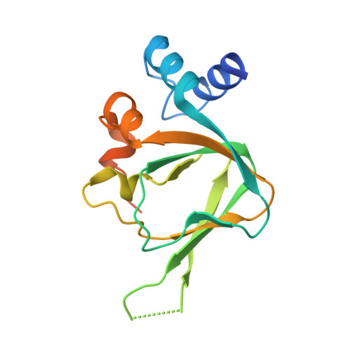
Joint X-ray/neutron structure of PKGIbeta in complex with cGMP
- PDB DOI: https://doi.org/10.2210/pdb4QXK/pdb
- Classification: SIGNALING PROTEIN
- Organism(s): Homo sapiens
- Expression System: Escherichia coli
- Mutation(s): No
- Deposited: 2014-07-21 Released: 2014-11-12
- Deposition Author(s): Kim, C., Gerlits, O., Kovalevsky, A., Huang, G.Y.
Experimental Data Snapshot
- Method: X-RAY DIFFRACTION
- R-Value Free: 0.260 (DCC)
- R-Value Work: 0.250 (DCC)
- Method: NEUTRON DIFFRACTION
- Resolution: 2.20 Å
- R-Value Free: 0.318 (Depositor)
- R-Value Work: 0.277 (Depositor)
- R-Value Observed: 0.277 (Depositor)
- 👁 Image
Download Mendeley
Neutron Diffraction Reveals Hydrogen Bonds Critical for cGMP-Selective Activation: Insights for cGMP-Dependent Protein Kinase Agonist Design.
Huang, G.Y., Gerlits, O.O., Blakeley, M.P., Sankaran, B., Kovalevsky, A.Y., Kim, C.(2014) Biochemistry 53: 6725-6727
- PubMed: 25271401 Search on PubMedSearch on PubMed Central
- DOI: https://doi.org/10.1021/bi501012v
- Primary Citation Related Structures:
4QX5, 4QXK - PubMed Abstract:
High selectivity of cyclic-nucleotide binding (CNB) domains for cAMP and cGMP are required for segregating signaling pathways; however, the mechanism of selectivity remains unclear. To investigate the mechanism of high selectivity in cGMP-dependent protein kinase (PKG), we determined a room-temperature joint X-ray/neutron (XN) structure of PKG Iβ CNB-B, a domain 200-fold selective for cGMP over cAMP, bound to cGMP (2.2 Å), and a low-temperature X-ray structure of CNB-B with cAMP (1.3 Å). The XN structure directly describes the hydrogen bonding interactions that modulate high selectivity for cGMP, while the structure with cAMP reveals that all these contacts are disrupted, explaining its low affinity for cAMP.
- Verna and Mars McClean Department of Biochemistry and Molecular Biology, Baylor College of Medicine , One Baylor Plaza, Houston, Texas 77004, United States.
Organizational Affiliation:
Explore in 3D: Structure | Sequence Annotations | Electron Density | Validation Report | Ligand Interaction (PCG)
Explore in 3D: Structure | Sequence Annotations | Electron Density | Validation Report | Ligand Interaction (PCG)
Global Symmetry: Asymmetric - C1
Global Stoichiometry: Monomer - A1
Find Similar Assemblies
Biological assembly 1 assigned by authors and generated by PISA (software)
Macromolecule Content
- Total Structure Weight: 17.31 kDa
- Atom Count: 1,138
- Modeled Residue Count: 127
- Deposited Residue Count: 153
- Unique protein chains: 1
Entity ID: 1 | |||||
|---|---|---|---|---|---|
| Molecule | Chains | Sequence Length | Organism | Details | Image |
| cGMP-dependent protein kinase 1 | 153 | Homo sapiens | Mutation(s): 0 Gene Names: PRKG1, PRKG1B, PRKGR1A, PRKGR1B EC: 2.7.11.12 | 👁 Image | |
UniProt & NIH Common Fund Data Resources | |||||
Find proteins for Q13976 (Homo sapiens) Explore Q13976 Go to UniProtKB: Q13976 | |||||
PHAROS: Q13976 GTEx: ENSG00000185532 | |||||
Entity Groups | |||||
| Sequence Clusters | 30% Identity50% Identity70% Identity90% Identity95% Identity100% Identity | ||||
| UniProt Group | Q13976 | ||||
Sequence AnnotationsExpand | |||||
| |||||
| Ligands 2 Unique | |||||
|---|---|---|---|---|---|
| ID | Chains | Name / Formula / InChI Key | 2D Diagram | 3D Interactions | |
| PCG Query on PCG Download Ideal Coordinates CCD File | B [auth A] | CYCLIC GUANOSINE MONOPHOSPHATE C10 H12 N5 O7 P ZOOGRGPOEVQQDX-UUOKFMHZSA-N | 👁 Image | ||
| NA Query on NA Download Ideal Coordinates CCD File | C [auth A] | SODIUM ION Na FKNQFGJONOIPTF-UHFFFAOYSA-N | 👁 Image | ||
Experimental Data
- Method: X-RAY DIFFRACTION
- R-Value Free: 0.260 (DCC)
- R-Value Work: 0.250 (DCC)
- Method: NEUTRON DIFFRACTION
- Resolution: 2.20 Å
- R-Value Free: 0.318 (Depositor)
- R-Value Work: 0.277 (Depositor)
- R-Value Observed: 0.277 (Depositor)
| Length ( Å ) | Angle ( ˚ ) |
|---|---|
| a = 48.746 | α = 90 |
| b = 48.746 | β = 90 |
| c = 104.861 | γ = 90 |
| Software Name | Purpose |
|---|---|
| nCNS | refinement |
| Maatel | data collection |
| HKL-3000 | data reduction |
| LAUEGEN | data reduction |
| HKL-3000 | data scaling |
| LSCALE | data scaling |
| CNS | phasing |
Deposition Data
- Released Date: 2014-11-12 Deposition Author(s): Kim, C., Gerlits, O., Kovalevsky, A., Huang, G.Y.
Revision History (Full details and data files)
- Version 1.0: 2014-11-12
Type: Initial release - Version 1.1: 2018-06-20
Changes: Data collection - Version 1.2: 2024-02-28
Changes: Data collection, Database references, Derived calculations