
- FASTA Sequence
- PDBx/mmCIF Format
- PDBx/mmCIF Format (gz)
- BinaryCIF Format (gz)
- Legacy PDB Format
- Legacy PDB Format (gz)
- PDBML/XML Format (gz)
- Structure Factors (CIF)
- Structure Factors (CIF - gz)
- Validation Full (PDF - gz)
- Validation (XML - gz)
- Validation (CIF - gz)
- Validation 2fo-fc coefficients (CIF - gz)
- Validation fo-fc coefficients (CIF - gz)
- Biological Assembly 1 (CIF - gz)
- Biological Assembly 1 (PDB - gz)
4TUU | pdb_00004tuu
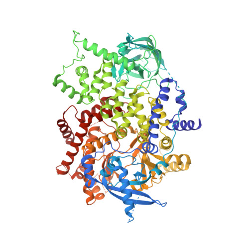
Isolated p110a subunit of PI3Ka provides a platform for structure-based drug design
- PDB DOI: https://doi.org/10.2210/pdb4TUU/pdb
- Classification: TRANSFERASE
- Organism(s): Homo sapiens
- Expression System: unidentified baculovirus
- Mutation(s): No
- Deposited: 2014-06-24 Released: 2014-08-06
- Deposition Author(s): Chen, P., Deng, Y.-L., Bergqvist, S., Falk, M., Liu, W., Timofeevski, S.
Experimental Data Snapshot
- Method: X-RAY DIFFRACTION
- Resolution: 2.64 Å
- R-Value Free: 0.245 (Depositor), 0.260 (DCC)
- R-Value Work: 0.209 (Depositor), 0.220 (DCC)
- R-Value Observed: 0.211 (Depositor)
wwPDB Validation 3D Report Full Report
- 👁 Image
Download Mendeley
Engineering of an isolated p110 alpha subunit of PI3K alpha permits crystallization and provides a platform for structure-based drug design.
Chen, P., Deng, Y.L., Bergqvist, S., Falk, M.D., Liu, W., Timofeevski, S., Brooun, A.(2014) Protein Sci 23: 1332-1340
- PubMed: 25043846 Search on PubMedSearch on PubMed Central
- DOI: https://doi.org/10.1002/pro.2517
- Primary Citation Related Structures:
4TUU, 4TV3 - PubMed Abstract:
PI3Kα remains an attractive target for the development of anticancer targeted therapy. A number of p110α crystal structures in complex with the nSH2-iSH2 fragment of p85 regulatory subunit have been reported, including a few small molecule co-crystal structures, but the utilization of this crystal form is limited by low diffraction resolution and a crystal packing artifact that partially blocks the ATP binding site. Taking advantage of recent data on the functional characterization of the lipid binding properties of p110α, we designed a set of novel constructs allowing production of isolated stable p110α subunit missing the Adapter Binding Domain and lacking or featuring a modified C-terminal lipid binding motif. While this protein is not catalytically competent to phosphorylate its substrate PIP2, it retains ligand binding properties as indicated by direct binding studies with a pan-PI3Kα inhibitor. Additionally, we determined apo and PF-04691502 bound crystal structures of the p110α (105-1048) subunit at 2.65 and 2.85 Å, respectively. Comparison of isolated p110α(105-1048) with the p110α/p85 complex reveals a high degree of structural similarity, which validates suitability of this catalytically inactive p110α for iterative SBDD. Importantly, this crystal form of p110α readily accommodates the binding of noncovalent inhibitor by means of a fully accessible ATP site. The strategy presented here can be also applied to structural studies of other members of PI3KIA family.
- Oncology Structural Biology, Worldwide Research and Development, Pfizer Inc., San Diego, California, 92121.
Organizational Affiliation:
Explore in 3D: Structure | Sequence Annotations | Electron Density | Validation Report
Explore in 3D: Structure | Sequence Annotations | Electron Density | Validation Report
Global Symmetry: Asymmetric - C1
Global Stoichiometry: Monomer - A1
Find Similar Assemblies
Biological assembly 1 assigned by authors and generated by PISA (software)
Macromolecule Content
- Total Structure Weight: 109.98 kDa
- Atom Count: 6,631
- Modeled Residue Count: 879
- Deposited Residue Count: 946
- Unique protein chains: 1
Entity ID: 1 | |||||
|---|---|---|---|---|---|
| Molecule | Chains | Sequence Length | Organism | Details | Image |
| Phosphatidylinositol 4,5-bisphosphate 3-kinase catalytic subunit alpha isoform | 946 | Homo sapiens | Mutation(s): 0 Gene Names: PIK3CA EC: 2.7.1.153 (PDB Primary Data), 2.7.11.1 (PDB Primary Data), 2.7.1.137 (UniProt) | 👁 Image | |
UniProt & NIH Common Fund Data Resources | |||||
Find proteins for P42336 (Homo sapiens) Explore P42336 Go to UniProtKB: P42336 | |||||
PHAROS: P42336 GTEx: ENSG00000121879 | |||||
Entity Groups | |||||
| Sequence Clusters | 30% Identity50% Identity70% Identity90% Identity95% Identity100% Identity | ||||
| UniProt Group | P42336 | ||||
Sequence AnnotationsExpand | |||||
| |||||
Experimental Data
- Method: X-RAY DIFFRACTION
- Resolution: 2.64 Å
- R-Value Free: 0.245 (Depositor), 0.260 (DCC)
- R-Value Work: 0.209 (Depositor), 0.220 (DCC)
- R-Value Observed: 0.211 (Depositor)
| Length ( Å ) | Angle ( ˚ ) |
|---|---|
| a = 57.689 | α = 90 |
| b = 136.447 | β = 90 |
| c = 143.083 | γ = 90 |
| Software Name | Purpose |
|---|---|
| BUSTER | refinement |
Deposition Data
- Released Date: 2014-08-06 Deposition Author(s): Chen, P., Deng, Y.-L., Bergqvist, S., Falk, M., Liu, W., Timofeevski, S.
Revision History (Full details and data files)
- Version 1.0: 2014-08-06
Type: Initial release - Version 1.1: 2014-10-01
Changes: Database references - Version 1.2: 2015-02-04
Changes: Derived calculations - Version 1.3: 2023-12-27
Changes: Data collection, Database references, Derived calculations, Other, Refinement description, Source and taxonomy