
- FASTA Sequence
- PDBx/mmCIF Format
- PDBx/mmCIF Format (gz)
- BinaryCIF Format (gz)
- Legacy PDB Format
- Legacy PDB Format (gz)
- PDBML/XML Format (gz)
- Structure Factors (CIF)
- Structure Factors (CIF - gz)
- Validation Full (PDF - gz)
- Validation (XML - gz)
- Validation (CIF - gz)
- Validation 2fo-fc coefficients (CIF - gz)
- Validation fo-fc coefficients (CIF - gz)
- Biological Assembly 1 (CIF - gz)
- Biological Assembly 1 (PDB - gz)
4YV8 | pdb_00004yv8
Crystal structure of cathepsin K bound to the covalent inhibitor lichostatinal
- PDB DOI: https://doi.org/10.2210/pdb4YV8/pdb
- Classification: HYDROLASE/INHIBITOR
- Organism(s): Homo sapiens, actinomycete 095-35
- Expression System: Pichia
- Mutation(s): No
- Deposited: 2015-03-19 Released: 2016-05-04
- Deposition Author(s): Aguda, A.H., Nguyen, N.T., Bromme, D., Brayer, G.D.
- Funding Organization(s): Canadian Institutes of Health Research (CIHR)
Experimental Data Snapshot
- Method: X-RAY DIFFRACTION
- Resolution: 2.00 Å
- R-Value Free: 0.205 (Depositor), 0.195 (DCC)
- R-Value Work: 0.173 (Depositor), 0.181 (DCC)
- R-Value Observed: 0.175 (Depositor)
Starting Model: experimental
View more details
wwPDB Validation3D Report Full Report
Literature
- 👁 Image
Download Mendeley
Affinity Crystallography: A New Approach to Extracting High-Affinity Enzyme Inhibitors from Natural Extracts.
Aguda, A.H., Lavallee, V., Cheng, P., Bott, T.M., Meimetis, L.G., Law, S., Nguyen, N.T., Williams, D.E., Kaleta, J., Villanueva, I., Davies, J., Andersen, R.J., Brayer, G.D., Bromme, D.(2016) J Nat Prod 79: 1962-1970
- PubMed: 27498895 Search on PubMed
- DOI: https://doi.org/10.1021/acs.jnatprod.6b00215
- Primary Citation Related Structures:
4YV8, 4YVA - PubMed Abstract:
Natural products are an important source of novel drug scaffolds. The highly variable and unpredictable timelines associated with isolating novel compounds and elucidating their structures have led to the demise of exploring natural product extract libraries in drug discovery programs. Here we introduce affinity crystallography as a new methodology that significantly shortens the time of the hit to active structure cycle in bioactive natural product discovery research. This affinity crystallography approach is illustrated by using semipure fractions of an actinomycetes culture extract to isolate and identify a cathepsin K inhibitor and to compare the outcome with the traditional assay-guided purification/structural analysis approach. The traditional approach resulted in the identification of the known inhibitor antipain (1) and its new but lower potency dehydration product 2, while the affinity crystallography approach led to the identification of a new high-affinity inhibitor named lichostatinal (3). The structure and potency of lichostatinal (3) was verified by total synthesis and kinetic characterization. To the best of our knowledge, this is the first example of isolating and characterizing a potent enzyme inhibitor from a partially purified crude natural product extract using a protein crystallographic approach.
- Department of Oral Biological and Medical Sciences, Faculty of Dentistry, ‡Department of Biochemistry and Molecular Biology, Faculty of Medicine, §Department of Chemistry and Earth, Ocean & Atmospheric Sciences, Faculty of Science, ⊥Department of Microbiology, Faculty of Science, and ∥Centre for Blood Research, University of British Columbia , Vancouver, BC Canada , V6T 1Z3.
Organizational Affiliation:
Explore in 3D: Structure | Sequence Annotations | Electron Density | Validation Report | Ligand Interaction (SO4)
Biological Assembly 1
Explore in 3D: Structure | Sequence Annotations | Electron Density | Validation Report | Ligand Interaction (SO4)
Global Symmetry: Asymmetric - C1
Global Stoichiometry: Hetero 2-mer - A1B1
Find Similar Assemblies
Biological assembly 1 assigned by authors and generated by PISA (software)
Macromolecule Content
- Total Structure Weight: 24.31 kDa
- Atom Count: 1,855
- Modeled Residue Count: 218
- Deposited Residue Count: 220
- Unique protein chains: 2
Macromolecules
Entity ID: 1 | |||||
|---|---|---|---|---|---|
| Molecule | Chains | Sequence Length | Organism | Details | Image |
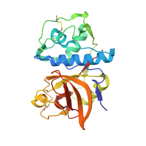
| Cathepsin K | 215 | Homo sapiens | Mutation(s): 0 Gene Names: CTSK, CTSO, CTSO2 EC: 3.4.22.38 | 👁 Image | |
UniProt & NIH Common Fund Data Resources | |||||
PHAROS: P43235 GTEx: ENSG00000143387 | |||||
Entity Groups | |||||
| Sequence Clusters | 30% Identity50% Identity70% Identity90% Identity95% Identity100% Identity | ||||
| UniProt Group | P43235 | ||||
Sequence AnnotationsExpand | |||||
Reference Sequence | |||||
Entity ID: 2 | |||||
|---|---|---|---|---|---|
| Molecule | Chains | Sequence Length | Organism | Details | Image |
| Lichostatinal | 5 | actinomycete 095-35 | Mutation(s): 0 | 👁 Image | |
Small Molecules
| Ligands 1 Unique | |||||
|---|---|---|---|---|---|
| ID | Chains | Name / Formula / InChI Key | 2D Diagram | 3D Interactions | |
| SO4 Download:Ideal Coordinates CCD File | C [auth A], D [auth A], E [auth A] | SULFATE ION O4 S QAOWNCQODCNURD-UHFFFAOYSA-L | 👁 Image | ||
| Modified Residues 1 Unique | |||||
|---|---|---|---|---|---|
| ID | Chains | Type | Formula | 2D Diagram | Parent |
| RGL Query on RGL | B | L-PEPTIDE LINKING | C6 H15 N4 O | 👁 Image | ARG |
Biologically Interesting Molecules (External Reference)
1 UniqueExperimental Data & Validation
Experimental Data
- Method: X-RAY DIFFRACTION
- Resolution: 2.00 Å
- R-Value Free: 0.205 (Depositor), 0.195 (DCC)
- R-Value Work: 0.173 (Depositor), 0.181 (DCC)
- R-Value Observed: 0.175 (Depositor)
| Length ( Å ) | Angle ( ˚ ) |
|---|---|
| a = 56.717 | α = 90 |
| b = 56.717 | β = 90 |
| c = 130.365 | γ = 90 |
| Software Name | Purpose |
|---|---|
| PHENIX | refinement |
| MOSFLM | data reduction |
| SCALA | data scaling |
| PHASER | phasing |
Entry History
& Funding InformationDeposition Data
- Released Date: 2016-05-04 Deposition Author(s): Aguda, A.H., Nguyen, N.T., Bromme, D., Brayer, G.D.
| Funding Organization | Location | Grant Number |
|---|---|---|
| Canadian Institutes of Health Research (CIHR) | Canada | FRN 111082 |
Revision History (Full details and data files)
- Version 1.0: 2016-05-04
Type: Initial release - Version 1.1: 2016-08-24
Changes: Database references - Version 1.2: 2016-09-07
Changes: Database references - Version 1.3: 2017-09-27
Changes: Data collection, Derived calculations - Version 2.0: 2019-05-15
Changes: Data collection, Polymer sequence - Version 2.1: 2020-01-08
Changes: Author supporting evidence - Version 2.2: 2023-09-27
Changes: Data collection, Database references, Refinement description - Version 2.3: 2023-11-15
Changes: Data collection