
- FASTA Sequence
- PDBx/mmCIF Format
- PDBx/mmCIF Format (gz)
- BinaryCIF Format (gz)
- Legacy PDB Format
- Legacy PDB Format (gz)
- PDBML/XML Format (gz)
- Structure Factors (CIF)
- Structure Factors (CIF - gz)
- Validation Full (PDF - gz)
- Validation (XML - gz)
- Validation (CIF - gz)
- Validation 2fo-fc coefficients (CIF - gz)
- Validation fo-fc coefficients (CIF - gz)
- Biological Assembly 1 (CIF - gz)
- Biological Assembly 1 (PDB - gz)
4ZA0 | pdb_00004za0
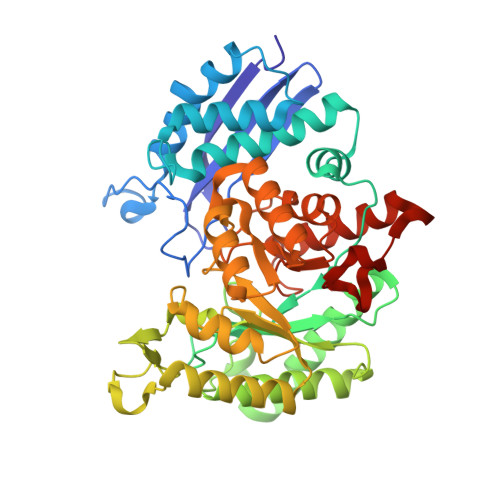
Structure of Human Enolase 2 in complex with Phosphonoacetohydroxamate
- PDB DOI: https://doi.org/10.2210/pdb4ZA0/pdb
- Classification: LYASE/LYASE INHIBITOR
- Organism(s): Homo sapiens
- Expression System: Escherichia coli BL21
- Mutation(s): No
- Deposited: 2015-04-13 Released: 2016-04-13
- Deposition Author(s): Leonard, P.G., Maxwell, D., Czako, B., Muller, F.L.
Experimental Data Snapshot
- Method: X-RAY DIFFRACTION
- Resolution: 2.31 Å
- R-Value Free: 0.204 (Depositor), 0.200 (DCC)
- R-Value Work: 0.164 (Depositor), 0.170 (DCC)
- R-Value Observed: 0.166 (Depositor)
Starting Model: experimental
View more details
- 👁 Image
Download Mendeley
SF2312 is a natural phosphonate inhibitor of enolase.
Leonard, P.G., Satani, N., Maxwell, D., Lin, Y.H., Hammoudi, N., Peng, Z., Pisaneschi, F., Link, T.M., Lee, G.R., Sun, D., Prasad, B.A., Di Francesco, M.E., Czako, B., Asara, J.M., Wang, Y.A., Bornmann, W., DePinho, R.A., Muller, F.L.(2016) Nat Chem Biol 12: 1053-1058
- PubMed: 27723749 Search on PubMedSearch on PubMed Central
- DOI: https://doi.org/10.1038/nchembio.2195
- Primary Citation Related Structures:
4ZA0, 4ZCW - PubMed Abstract:
Despite being crucial for energy generation in most forms of life, few if any microbial antibiotics specifically inhibit glycolysis. To develop a specific inhibitor of the glycolytic enzyme enolase 2 (ENO2) for the treatment of cancers with deletion of ENO1 (encoding enolase 1), we modeled the synthetic tool compound inhibitor phosphonoacetohydroxamate (PhAH) into the active site of human ENO2. A ring-stabilized analog of PhAH, in which the hydroxamic nitrogen is linked to Cα by an ethylene bridge, was predicted to increase binding affinity by stabilizing the inhibitor in a bound conformation. Unexpectedly, a structure-based search revealed that our hypothesized backbone-stabilized PhAH bears strong similarity to SF2312, a phosphonate antibiotic of unknown mode of action produced by the actinomycete Micromonospora, which is active under anaerobic conditions. Here, we present multiple lines of evidence, including a novel X-ray structure, that SF2312 is a highly potent, low-nanomolar inhibitor of enolase.
- Department of Genomic Medicine and Core for Biomolecular Structure and Function, University of Texas MD Anderson Cancer Center, Houston, TX 77054.
- Department of Cancer Systems Imaging, University of Texas MD Anderson Cancer Center, Houston, TX 77054.
- Department of Clinical Analytics & Informatics, Houston, TX 77054-3403.
- Cardtronics, Inc., Houston, TX 77042.
- Institute for Applied Cancer Science, University of Texas MD Anderson Cancer Center, Houston, TX 77054.
- Department of Medicine, Beth Israel Deaconess Medical Center and Harvard Medical School, Boston, MA 02115.
- Department of Cancer Biology, University of Texas MD Anderson Cancer Center, Houston, TX 77030, USA University of Texas MD Anderson Cancer Center, Houston, TX 77054 USA.
- Bayou Therapeutics, Inc, Missouri City, TX 77459-3028.
Organizational Affiliation:
Explore in 3D: Structure | Sequence Annotations | Electron Density | Validation Report | Ligand Interaction (PAH)
Explore in 3D: Structure | Sequence Annotations | Electron Density | Validation Report | Ligand Interaction (PAH)
Global Symmetry: Cyclic - C2 (Explore in 3D)
Global Stoichiometry: Homo 2-mer - A2
Find Similar Assemblies
Biological assembly 1 assigned by authors and generated by PISA (software)
Macromolecule Content
- Total Structure Weight: 96.71 kDa
- Atom Count: 7,139
- Modeled Residue Count: 866
- Deposited Residue Count: 880
- Unique protein chains: 1
Entity ID: 1 | |||||
|---|---|---|---|---|---|
| Molecule | Chains | Sequence Length | Organism | Details | Image |
| Gamma-enolase | 440 | Homo sapiens | Mutation(s): 0 Gene Names: ENO2 EC: 4.2.1.11 | 👁 Image | |
UniProt & NIH Common Fund Data Resources | |||||
Find proteins for P09104 (Homo sapiens) Explore P09104 Go to UniProtKB: P09104 | |||||
PHAROS: P09104 GTEx: ENSG00000111674 | |||||
Entity Groups | |||||
| Sequence Clusters | 30% Identity50% Identity70% Identity90% Identity95% Identity100% Identity | ||||
| UniProt Group | P09104 | ||||
Sequence AnnotationsExpand | |||||
| |||||
| Ligands 2 Unique | |||||
|---|---|---|---|---|---|
| ID | Chains | Name / Formula / InChI Key | 2D Diagram | 3D Interactions | |
| PAH Query on PAH Download Ideal Coordinates CCD File | E [auth A], H [auth B] | PHOSPHONOACETOHYDROXAMIC ACID C2 H6 N O5 P LDKRAXXVBWHMRH-UHFFFAOYSA-N | 👁 Image | ||
| MG Query on MG Download Ideal Coordinates CCD File
| C [auth A], D [auth A], F [auth B], G [auth B] | MAGNESIUM ION Mg JLVVSXFLKOJNIY-UHFFFAOYSA-N | 👁 Image | ||
Experimental Data
- Method: X-RAY DIFFRACTION
- Resolution: 2.31 Å
- R-Value Free: 0.204 (Depositor), 0.200 (DCC)
- R-Value Work: 0.164 (Depositor), 0.170 (DCC)
- R-Value Observed: 0.166 (Depositor)
| Length ( Å ) | Angle ( ˚ ) |
|---|---|
| a = 67.79 | α = 90 |
| b = 108.671 | β = 90 |
| c = 112.21 | γ = 90 |
| Software Name | Purpose |
|---|---|
| iMOSFLM | data reduction |
| Aimless | data scaling |
| PHASER | phasing |
| PHENIX | refinement |
| Coot | model building |
| PDB_EXTRACT | data extraction |
Deposition Data
- Released Date: 2016-04-13 Deposition Author(s): Leonard, P.G., Maxwell, D., Czako, B., Muller, F.L.
Revision History (Full details and data files)
- Version 1.0: 2016-04-13
Type: Initial release - Version 1.1: 2016-10-05
Changes: Database references - Version 1.2: 2016-10-26
Changes: Database references - Version 1.3: 2016-11-23
Changes: Database references - Version 1.4: 2017-11-22
Changes: Refinement description - Version 1.5: 2023-09-27
Changes: Data collection, Database references, Derived calculations, Refinement description