
- FASTA Sequence
- PDBx/mmCIF Format
- PDBx/mmCIF Format (gz)
- BinaryCIF Format (gz)
- Legacy PDB Format
- Legacy PDB Format (gz)
- PDBML/XML Format (gz)
- Structure Factors (CIF)
- Structure Factors (CIF - gz)
- Validation Full (PDF - gz)
- Validation (XML - gz)
- Validation (CIF - gz)
- Validation 2fo-fc coefficients (CIF - gz)
- Validation fo-fc coefficients (CIF - gz)
- Biological Assembly 1 (CIF - gz)
- Biological Assembly 1 (PDB - gz)
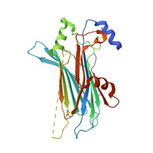
4ZKB | pdb_00004zkb
The chemokine binding protein of orf virus complexed with CCL3
- PDB DOI: https://doi.org/10.2210/pdb4ZKB/pdb
- Classification: Viral Protein/cytokine
- Organism(s): Orf virus (strain NZ2), Homo sapiens
- Expression System: Homo sapiens
- Mutation(s): No
- Deposited: 2015-04-30 Released: 2015-07-08
- Deposition Author(s): Knapp, K.M., Nakatani, Y., Krause, K.L.
Experimental Data Snapshot
- Method: X-RAY DIFFRACTION
- Resolution: 2.90 Å
- R-Value Free: 0.323 (Depositor), 0.330 (DCC)
- R-Value Work: 0.268 (Depositor), 0.270 (DCC)
- R-Value Observed: 0.270 (Depositor)
Starting Model: experimental
View more details
wwPDB Validation 3D Report Full Report
- 👁 Image
Download Mendeley
Structures of Orf Virus Chemokine Binding Protein in Complex with Host Chemokines Reveal Clues to Broad Binding Specificity.
Counago, R.M., Knapp, K.M., Nakatani, Y., Fleming, S.B., Corbett, M., Wise, L.M., Mercer, A.A., Krause, K.L.(2015) Structure 23: 1199-1213
- PubMed: 26095031 Search on PubMed
- DOI: https://doi.org/10.1016/j.str.2015.04.023
- Primary Citation Related Structures:
4P5I, 4ZK9, 4ZKB, 4ZKC - PubMed Abstract:
The chemokine binding protein (CKBP) from orf virus (ORFV) binds with high affinity to chemokines from three classes, C, CC, and CXC, making it unique among poxvirus CKBPs described to date. We present its crystal structure alone and in complex with three CC chemokines, CCL2, CCL3, and CCL7. ORFV CKBP possesses a β-sandwich fold that is electrostatically and sterically complementary to its binding partners. Chemokines bind primarily through interactions involving the N-terminal loop and a hydrophobic recess on the ORFV CKBP β-sheet II surface, and largely polar interactions between the chemokine 20s loop and a negatively charged surface groove located at one end of the CKBP β-sheet II surface. ORFV CKBP interacts with leukocyte receptor and glycosaminoglycan binding sites found on the surface of bound chemokines. SEC-MALLS and chromatographic evidence is presented supporting that ORFV CKBP is a dimer in solution over a broad range of protein concentrations.
- Department of Biochemistry, University of Otago, Dunedin 9054, New Zealand.
- Department of Biochemistry, University of Otago, Dunedin 9054, New Zealand; School of Biological Sciences, Bangor University, Bangor LL57 2UW, UK.
- Department of Biochemistry, University of Otago, Dunedin 9054, New Zealand; Department of Microbiology and Immunology, University of Otago, Dunedin 9054, New Zealand.
- Department of Microbiology and Immunology, University of Otago, Dunedin 9054, New Zealand.
- Department of Biochemistry, University of Otago, Dunedin 9054, New Zealand. Electronic address: kurt.krause@otago.ac.nz.
Organizational Affiliation:
Explore in 3D: Structure | Sequence Annotations | Electron Density | Validation Report
Explore in 3D: Structure | Sequence Annotations | Electron Density | Validation Report
Global Symmetry: Asymmetric - C1
Global Stoichiometry: Hetero 2-mer - A1B1
Find Similar Assemblies
Biological assembly 1 assigned by authors and generated by PISA (software)
Macromolecule Content
- Total Structure Weight: 39.94 kDa
- Atom Count: 1,919
- Modeled Residue Count: 267
- Deposited Residue Count: 351
- Unique protein chains: 2
Entity ID: 1 | |||||
|---|---|---|---|---|---|
| Molecule | Chains | Sequence Length | Organism | Details | Image |
| Chemokine binding protein | 276 | Orf virus (strain NZ2) | Mutation(s): 0 | 👁 Image | |
UniProt | |||||
Find proteins for Q2F862 (Orf virus (strain NZ2)) Explore Q2F862 Go to UniProtKB: Q2F862 | |||||
Entity Groups | |||||
| Sequence Clusters | 30% Identity50% Identity70% Identity90% Identity95% Identity100% Identity | ||||
| UniProt Group | Q2F862 | ||||
Glycosylation | |||||
| Glycosylation Sites: 2 | |||||
Sequence AnnotationsExpand | |||||
| |||||
Entity ID: 2 | |||||
|---|---|---|---|---|---|
| Molecule | Chains | Sequence Length | Organism | Details | Image |
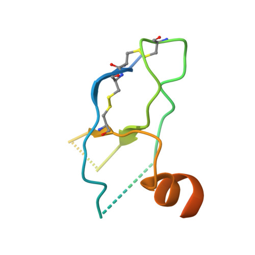
| C-C motif chemokine 3 | 75 | Homo sapiens | Mutation(s): 0 Gene Names: CCL3, G0S19-1, MIP1A, SCYA3 | 👁 Image | |
UniProt & NIH Common Fund Data Resources | |||||
Find proteins for P10147 (Homo sapiens) Explore P10147 Go to UniProtKB: P10147 | |||||
PHAROS: P10147 GTEx: ENSG00000277632 | |||||
Entity Groups | |||||
| Sequence Clusters | 30% Identity50% Identity70% Identity90% Identity95% Identity100% Identity | ||||
| UniProt Group | P10147 | ||||
Sequence AnnotationsExpand | |||||
| |||||
Entity ID: 3 | |||||
|---|---|---|---|---|---|
| Molecule | Chains | Length | 2D Diagram | Glycosylation | 3D Interactions |
| 2-acetamido-2-deoxy-beta-D-glucopyranose-(1-4)-2-acetamido-2-deoxy-beta-D-glucopyranose | C | 2 | 👁 Image | N-Glycosylation | |
Glycosylation Resources | |||||
GlyTouCan: G42666HT GlyCosmos: G42666HT GlyGen: G42666HT | |||||
Experimental Data
- Method: X-RAY DIFFRACTION
- Resolution: 2.90 Å
- R-Value Free: 0.323 (Depositor), 0.330 (DCC)
- R-Value Work: 0.268 (Depositor), 0.270 (DCC)
- R-Value Observed: 0.270 (Depositor)
| Length ( Å ) | Angle ( ˚ ) |
|---|---|
| a = 78 | α = 90 |
| b = 78 | β = 90 |
| c = 186.02 | γ = 90 |
| Software Name | Purpose |
|---|---|
| REFMAC | refinement |
| XDS | data reduction |
| SCALA | data scaling |
| PHASER | phasing |
Deposition Data
- Released Date: 2015-07-08 Deposition Author(s): Knapp, K.M., Nakatani, Y., Krause, K.L.
Revision History (Full details and data files)
- Version 1.0: 2015-07-08
Type: Initial release - Version 1.1: 2015-07-15
Changes: Database references - Version 1.2: 2018-06-13
Changes: Data collection, Database references, Derived calculations, Source and taxonomy, Structure summary - Version 2.0: 2020-07-29
Type: Remediation
Reason: Carbohydrate remediation
Changes: Atomic model, Data collection, Derived calculations, Structure summary - Version 2.1: 2023-09-27
Changes: Data collection, Database references, Refinement description, Structure summary - Version 2.2: 2024-11-13
Changes: Structure summary