
- FASTA Sequence
- PDBx/mmCIF Format
- PDBx/mmCIF Format (gz)
- BinaryCIF Format (gz)
- Legacy PDB Format
- Legacy PDB Format (gz)
- PDBML/XML Format (gz)
- Structure Factors (CIF)
- Structure Factors (CIF - gz)
- Validation Full (PDF - gz)
- Validation (XML - gz)
- Validation (CIF - gz)
- Validation 2fo-fc coefficients (CIF - gz)
- Validation fo-fc coefficients (CIF - gz)
- Biological Assembly 1 (CIF - gz)
- Biological Assembly 2 (CIF - gz)
- Biological Assembly 3 (CIF - gz)
- Biological Assembly 4 (CIF - gz)
- Biological Assembly 1 (PDB - gz)
- Biological Assembly 2 (PDB - gz)
- Biological Assembly 3 (PDB - gz)
- Biological Assembly 4 (PDB - gz)
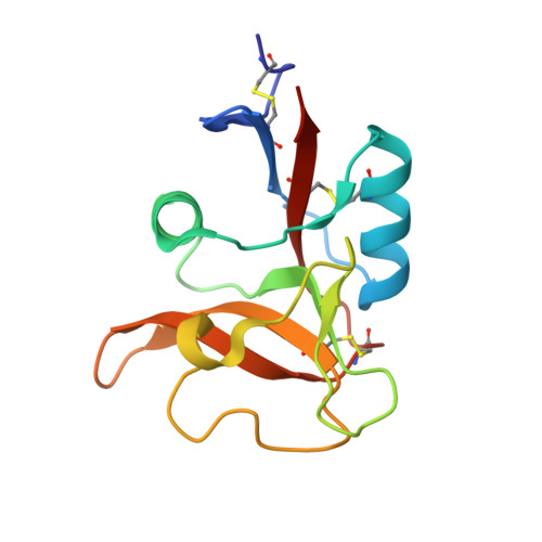
5B1W | pdb_00005b1w
Crystal structure of human dendritic cell inhibitory receptor (DCIR) C-type lectin domain in ligand-free form
- PDB DOI: https://doi.org/10.2210/pdb5B1W/pdb
- Classification: CARBOHYDRATE BINDING PROTEIN
- Organism(s): Homo sapiens
- Expression System: Escherichia coli
- Mutation(s): No
- Deposited: 2015-12-21 Released: 2016-05-11
- Deposition Author(s): Nagae, M., Yamaguchi, Y.
- Funding Organization(s): MEXT
Experimental Data Snapshot
- Method: X-RAY DIFFRACTION
- Resolution: 3.05 Å
- R-Value Free: 0.284 (Depositor), 0.278 (DCC)
- R-Value Work: 0.253 (Depositor), 0.256 (DCC)
- R-Value Observed: 0.254 (Depositor)
Starting Model: experimental
View more details
wwPDB Validation3D Report Full Report
Literature
- 👁 Image
Download Mendeley
Crystal structure of human dendritic cell inhibitory receptor C-type lectin domain reveals the binding mode with N-glycan
Nagae, M., Ikeda, A., Hanashima, S., Kojima, T., Matsumoto, N., Yamamoto, K., Yamaguchi, Y.(2016) FEBS Lett 590: 1280-1288
- PubMed: 27015765 Search on PubMed
- DOI: https://doi.org/10.1002/1873-3468.12162
- Primary Citation Related Structures:
5B1W, 5B1X - PubMed Abstract:
Human dendritic cell inhibitory receptor (DCIR) is a C-type lectin receptor expressed in classical dendritic cells and accepts several oligosaccharide ligands including N-glycans. Here, we report the crystal structures of human DCIR C-type lectin domains in the absence and presence of a branched N-glycan unit. The domain has a typical C-type lectin fold and two bound calcium ions. In the ligand-bound form, the disaccharide unit (GlcNAcβ1-2Man) acceptably fits the electron density map, indicating that it forms the main epitope. The recognition of the nonterminal N-glycan unit explains the relatively broad specificity of this lectin.
- Structural Glycobiology Team, Systems Glycobiology Research Group, RIKEN-Max Planck Joint Research Center, RIKEN Global Research Cluster, Wako, Saitama, Japan.
- Department of Chemistry, Osaka University, Machikaneyama, Toyonaka, Osaka, Japan.
- Department of Integrated Biosciences, Graduate School of Frontier Sciences, The University of Tokyo, Chiba, Japan.
Organizational Affiliation:
Explore in 3D: Structure | Sequence Annotations | Electron Density | Validation Report | Ligand Interaction (CA)
Biological Assembly 1
Explore in 3D: Structure | Sequence Annotations | Electron Density | Validation Report | Ligand Interaction (CA)
Global Symmetry: Asymmetric - C1
Global Stoichiometry: Monomer - A1
Find Similar Assemblies
Biological assembly 1 assigned by authors and generated by PISA (software)
Biological Assembly 2
Explore in 3D: Structure | Sequence Annotations | Electron Density | Validation Report | Ligand Interaction (CA)
Global Symmetry: Asymmetric - C1
Global Stoichiometry: Monomer - A1
Find Similar Assemblies
Biological assembly 2 assigned by authors and generated by PISA (software)
Biological Assembly 3
Explore in 3D: Structure | Sequence Annotations | Electron Density | Validation Report | Ligand Interaction (CA)
Global Symmetry: Asymmetric - C1
Global Stoichiometry: Monomer - A1
Find Similar Assemblies
Biological assembly 3 assigned by authors and generated by PISA (software)
Biological Assembly 4
Explore in 3D: Structure | Sequence Annotations | Electron Density | Validation Report | Ligand Interaction (CA)
Global Symmetry: Asymmetric - C1
Global Stoichiometry: Monomer - A1
Find Similar Assemblies
Biological assembly 4 assigned by authors and generated by PISA (software)
Macromolecule Content
- Total Structure Weight: 63.57 kDa
- Atom Count: 4,301
- Modeled Residue Count: 516
- Deposited Residue Count: 536
- Unique protein chains: 1
Macromolecules
Entity ID: 1 | |||||
|---|---|---|---|---|---|
| Molecule | Chains | Sequence Length | Organism | Details | Image |
| C-type lectin domain family 4 member A | 134 | Homo sapiens | Mutation(s): 0 Gene Names: CLEC4A, CLECSF6, DCIR, LLIR, HDCGC13P | 👁 Image | |
UniProt & NIH Common Fund Data Resources | |||||
PHAROS: Q9UMR7 GTEx: ENSG00000111729 | |||||
Entity Groups | |||||
| Sequence Clusters | 30% Identity50% Identity70% Identity90% Identity95% Identity100% Identity | ||||
| UniProt Group | Q9UMR7 | ||||
Sequence AnnotationsExpand | |||||
Reference Sequence | |||||
Small Molecules
| Ligands 1 Unique | |||||
|---|---|---|---|---|---|
| ID | Chains | Name / Formula / InChI Key | 2D Diagram | 3D Interactions | |
| CA Download:Ideal Coordinates CCD File
| E [auth A] F [auth A] G [auth B] H [auth B] I [auth C] E [auth A], F [auth A], G [auth B], H [auth B], I [auth C], J [auth C], K [auth D], L [auth D] | CALCIUM ION Ca BHPQYMZQTOCNFJ-UHFFFAOYSA-N | 👁 Image | ||
Experimental Data & Validation
Experimental Data
- Method: X-RAY DIFFRACTION
- Resolution: 3.05 Å
- R-Value Free: 0.284 (Depositor), 0.278 (DCC)
- R-Value Work: 0.253 (Depositor), 0.256 (DCC)
- R-Value Observed: 0.254 (Depositor)
| Length ( Å ) | Angle ( ˚ ) |
|---|---|
| a = 102.489 | α = 90 |
| b = 105.832 | β = 90 |
| c = 65.387 | γ = 90 |
| Software Name | Purpose |
|---|---|
| PHENIX | refinement |
| HKL-2000 | data reduction |
| HKL-2000 | data scaling |
| MOLREP | phasing |
Entry History
& Funding InformationDeposition Data
- Released Date: 2016-05-11 Deposition Author(s): Nagae, M., Yamaguchi, Y.
| Funding Organization | Location | Grant Number |
|---|---|---|
| MEXT | Japan | 24770111 |
| MEXT | Japan | 15K18496 |
Revision History (Full details and data files)
- Version 1.0: 2016-05-11
Type: Initial release - Version 1.1: 2020-02-26
Changes: Data collection, Database references, Derived calculations - Version 1.2: 2023-11-08
Changes: Data collection, Database references, Derived calculations, Refinement description - Version 1.3: 2024-11-13
Changes: Structure summary