
- FASTA Sequence
- PDBx/mmCIF Format
- PDBx/mmCIF Format (gz)
- BinaryCIF Format (gz)
- Legacy PDB Format
- Legacy PDB Format (gz)
- PDBML/XML Format (gz)
- Structure Factors (CIF)
- Structure Factors (CIF - gz)
- Validation Full (PDF - gz)
- Validation (XML - gz)
- Validation (CIF - gz)
- Validation 2fo-fc coefficients (CIF - gz)
- Validation fo-fc coefficients (CIF - gz)
- Biological Assembly 1 (CIF - gz)
- Biological Assembly 1 (PDB - gz)
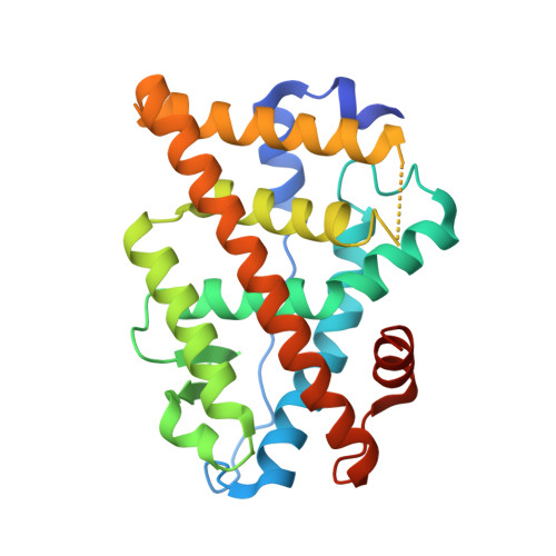
5E19 | pdb_00005e19
Crystal Structure of the ER-alpha Ligand-binding Domain in Complex with the Cyclofenil Derivative methyl {4-[bis(4-hydroxyphenyl)methylidene]cyclohexyl}acetate
- PDB DOI: https://doi.org/10.2210/pdb5E19/pdb
- Classification: TRANSCRIPTION
- Organism(s): Homo sapiens
- Expression System: Escherichia coli BL21(DE3)
- Mutation(s): Yes
- Deposited: 2015-09-29 Released: 2016-05-04
- Deposition Author(s): Nwachukwu, J.C., Srinivasan, S., Zheng, Y., Wang, S., Min, J., Dong, C., Liao, Z., Cavett, V., Nowak, J., Houtman, R., Carlson, K.E., Josan, J.S., Elemento, O., Katzenellenbogen, J.A., Zhou, H.B., Nettles, K.W.
Experimental Data Snapshot
- Method: X-RAY DIFFRACTION
- Resolution: 2.24 Å
- R-Value Free: 0.233 (Depositor), 0.230 (DCC)
- R-Value Work: 0.194 (Depositor), 0.200 (DCC)
- R-Value Observed: 0.197 (Depositor)
Starting Model: experimental
View more details
- 👁 Image
Download Mendeley
Predictive features of ligand-specific signaling through the estrogen receptor.
Nwachukwu, J.C., Srinivasan, S., Zheng, Y., Wang, S., Min, J., Dong, C., Liao, Z., Nowak, J., Wright, N.J., Houtman, R., Carlson, K.E., Josan, J.S., Elemento, O., Katzenellenbogen, J.A., Zhou, H.B., Nettles, K.W.(2016) Mol Syst Biol 12: 864-864
- PubMed: 27107013 Search on PubMedSearch on PubMed Central
- DOI: https://doi.org/10.15252/msb.20156701
- Primary Citation Related Structures:
4ZN7, 4ZNH, 4ZNS, 4ZNT, 4ZNU, 4ZNV, 4ZNW, 5DI7, 5DID, 5DIE, 5DIG, 5DK9, 5DKB, 5DKE, 5DKG, 5DKS, 5DL4, 5DLR, 5DMC, 5DMF, 5DP0, 5DRJ, 5DRM, 5DTV, 5DU5, 5DUE, 5DUG, 5DUH, 5DVS, 5DVV, 5DWE, 5DWG, 5DWI, 5DWJ, 5DXK, 5DXM, 5DXP, 5DXQ, 5DXR, 5DY8, 5DYB, 5DYD, 5DZ0, 5DZ1, 5DZ3, 5DZH, 5DZI, 5E0W, 5E0X, 5E14, ... Search all related entries - PubMed Abstract:
Some estrogen receptor-α (ERα)-targeted breast cancer therapies such as tamoxifen have tissue-selective or cell-specific activities, while others have similar activities in different cell types. To identify biophysical determinants of cell-specific signaling and breast cancer cell proliferation, we synthesized 241 ERα ligands based on 19 chemical scaffolds, and compared ligand response using quantitative bioassays for canonical ERα activities and X-ray crystallography. Ligands that regulate the dynamics and stability of the coactivator-binding site in the C-terminal ligand-binding domain, called activation function-2 (AF-2), showed similar activity profiles in different cell types. Such ligands induced breast cancer cell proliferation in a manner that was predicted by the canonical recruitment of the coactivators NCOA1/2/3 and induction of the GREB1 proliferative gene. For some ligand series, a single inter-atomic distance in the ligand-binding domain predicted their proliferative effects. In contrast, the N-terminal coactivator-binding site, activation function-1 (AF-1), determined cell-specific signaling induced by ligands that used alternate mechanisms to control cell proliferation. Thus, incorporating systems structural analyses with quantitative chemical biology reveals how ligands can achieve distinct allosteric signaling outcomes through ERα.
- Department of Cancer Biology, The Scripps Research Institute, Jupiter, FL, USA.
- State Key Laboratory of Virology, Key Laboratory of Combinatorial Biosynthesis and Drug Discovery (Wuhan University), Ministry of Education, Wuhan University School of Pharmaceutical Sciences, Wuhan, China.
- Department of Chemistry, University of Illinois, Urbana, IL, USA.
- PamGene International, Den Bosch, The Netherlands.
- Department of Chemistry, Virginia Tech, Blacksburg, VA, USA.
- Department of Physiology and Biophysics, Institute for Computational Biomedicine, Weill Cornell Medical College, New York, NY, USA.
- Department of Chemistry, University of Illinois, Urbana, IL, USA knettles@scripps.edu jkatzene@illinois.edu zhouhb@whu.edu.cn.
- State Key Laboratory of Virology, Key Laboratory of Combinatorial Biosynthesis and Drug Discovery (Wuhan University), Ministry of Education, Wuhan University School of Pharmaceutical Sciences, Wuhan, China knettles@scripps.edu jkatzene@illinois.edu zhouhb@whu.edu.cn.
- Department of Cancer Biology, The Scripps Research Institute, Jupiter, FL, USA knettles@scripps.edu jkatzene@illinois.edu zhouhb@whu.edu.cn.
Organizational Affiliation:
Explore in 3D: Structure | Sequence Annotations | Electron Density | Validation Report | Ligand Interaction (5K7)
Explore in 3D: Structure | Sequence Annotations | Electron Density | Validation Report | Ligand Interaction (5K7)
Global Symmetry: Cyclic - C2 (Explore in 3D)
Global Stoichiometry: Hetero 4-mer - A2B2
Pseudo Symmetry: Asymmetric - C1
Pseudo Stoichiometry: Hetero 4-mer - A2B1C1
Find Similar Assemblies
Biological assembly 1 assigned by authors and generated by PISA (software)
Macromolecule Content
- Total Structure Weight: 62.64 kDa
- Atom Count: 3,974
- Modeled Residue Count: 490
- Deposited Residue Count: 542
- Unique protein chains: 2
Entity ID: 1 | |||||
|---|---|---|---|---|---|
| Molecule | Chains | Sequence Length | Organism | Details | Image |
| Estrogen receptor | 257 | Homo sapiens | Mutation(s): 1 Gene Names: ESR1, ESR, NR3A1 | 👁 Image | |
UniProt & NIH Common Fund Data Resources | |||||
Find proteins for P03372 (Homo sapiens) Explore P03372 Go to UniProtKB: P03372 | |||||
PHAROS: P03372 GTEx: ENSG00000091831 | |||||
Entity Groups | |||||
| Sequence Clusters | 30% Identity50% Identity70% Identity90% Identity95% Identity100% Identity | ||||
| UniProt Group | P03372 | ||||
Sequence AnnotationsExpand | |||||
| |||||
Find similar proteins by: Sequence | 3D Structure
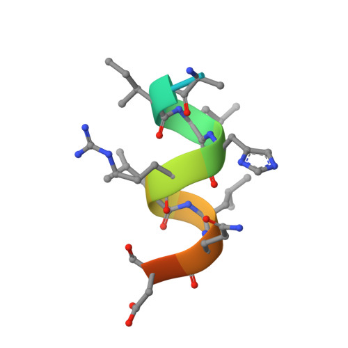
Entity ID: 2 | |||||
|---|---|---|---|---|---|
| Molecule | Chains | Sequence Length | Organism | Details | Image |
| Nuclear receptor coactivator 2 | 14 | Homo sapiens | Mutation(s): 0 | 👁 Image | |
UniProt & NIH Common Fund Data Resources | |||||
Find proteins for Q15596 (Homo sapiens) Explore Q15596 Go to UniProtKB: Q15596 | |||||
PHAROS: Q15596 GTEx: ENSG00000140396 | |||||
Entity Groups | |||||
| Sequence Clusters | 30% Identity50% Identity70% Identity90% Identity95% Identity100% Identity | ||||
| UniProt Group | Q15596 | ||||
Sequence AnnotationsExpand | |||||
| |||||
| Ligands 1 Unique | |||||
|---|---|---|---|---|---|
| ID | Chains | Name / Formula / InChI Key | 2D Diagram | 3D Interactions | |
| 5K7 Query on 5K7 Download Ideal Coordinates CCD File | E [auth A], F [auth B] | methyl {4-[bis(4-hydroxyphenyl)methylidene]cyclohexyl}acetate C22 H24 O4 ANBKAPQYIYCLKX-UHFFFAOYSA-N | 👁 Image | ||
Experimental Data
- Method: X-RAY DIFFRACTION
- Resolution: 2.24 Å
- R-Value Free: 0.233 (Depositor), 0.230 (DCC)
- R-Value Work: 0.194 (Depositor), 0.200 (DCC)
- R-Value Observed: 0.197 (Depositor)
| Length ( Å ) | Angle ( ˚ ) |
|---|---|
| a = 55.37 | α = 90 |
| b = 81.998 | β = 110.83 |
| c = 58.479 | γ = 90 |
| Software Name | Purpose |
|---|---|
| HKL-2000 | data scaling |
| PHENIX | refinement |
| PDB_EXTRACT | data extraction |
| HKL-2000 | data reduction |
| PHENIX | phasing |
Deposition Data
- Released Date: 2016-05-04 Deposition Author(s): Nwachukwu, J.C., Srinivasan, S., Zheng, Y., Wang, S., Min, J., Dong, C., Liao, Z., Cavett, V., Nowak, J., Houtman, R., Carlson, K.E., Josan, J.S., Elemento, O., Katzenellenbogen, J.A., Zhou, H.B., Nettles, K.W.
Revision History (Full details and data files)
- Version 1.0: 2016-05-04
Type: Initial release - Version 1.1: 2023-09-27
Changes: Data collection, Database references, Derived calculations, Refinement description