
- FASTA Sequence
- PDBx/mmCIF Format
- PDBx/mmCIF Format (gz)
- BinaryCIF Format (gz)
- Legacy PDB Format
- Legacy PDB Format (gz)
- PDBML/XML Format (gz)
- Structure Factors (CIF)
- Structure Factors (CIF - gz)
- Validation Full (PDF - gz)
- Validation (XML - gz)
- Validation (CIF - gz)
- Validation 2fo-fc coefficients (CIF - gz)
- Validation fo-fc coefficients (CIF - gz)
- Biological Assembly 1 (CIF - gz)
- Biological Assembly 2 (CIF - gz)
- Biological Assembly 3 (CIF - gz)
- Biological Assembly 4 (CIF - gz)
- Biological Assembly 1 (PDB - gz)
- Biological Assembly 2 (PDB - gz)
- Biological Assembly 3 (PDB - gz)
- Biological Assembly 4 (PDB - gz)
5ES4 | pdb_00005es4
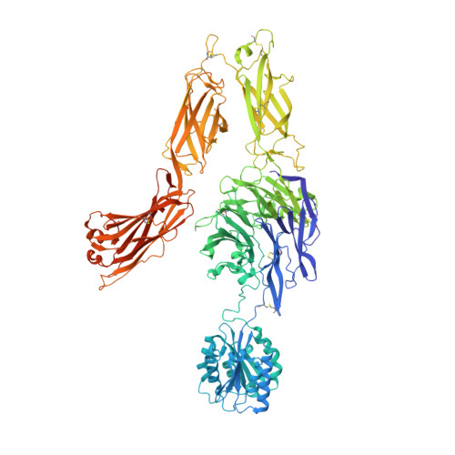
RE-REFINEMENT OF INTEGRIN ALPHAXBETA2 ECTODOMAIN IN THE CLOSED/BENT CONFORMATION
- PDB DOI: https://doi.org/10.2210/pdb5ES4/pdb
- Classification: CELL ADHESION
- Organism(s): Homo sapiens
- Expression System: Cricetulus griseus
- Mutation(s): No
- Deposited: 2015-11-16 Released: 2016-03-02
- Deposition Author(s): Sen, M., Springer, T.A.
- Funding Organization(s): National Institutes of Health/National Cancer Institute (NIH/NCI)
Experimental Data Snapshot
- Method: X-RAY DIFFRACTION
- Resolution: 3.30 Å
- R-Value Free: 0.307 (Depositor), 0.308 (DCC)
- R-Value Work: 0.257 (Depositor), 0.260 (DCC)
- R-Value Observed: 0.258 (Depositor)
Re-refinement Note
This entry reflects an alternative modeling of the original data in: 3k6s
Literature
- 👁 Image
Download Mendeley
Leukocyte integrin alpha L beta 2 headpiece structures: The alpha I domain, the pocket for the internal ligand, and concerted movements of its loops.
Sen, M., Springer, T.A.(2016) Proc Natl Acad Sci U S A 113: 2940-2945
- PubMed: 26936951 Search on PubMedSearch on PubMed Central
- DOI: https://doi.org/10.1073/pnas.1601379113
- Primary Citation Related Structures:
5E6R, 5E6S, 5E6U, 5E6V, 5E6W, 5E6X, 5ES4 - PubMed Abstract:
High-resolution crystal structures of the headpiece of lymphocyte function-associated antigen-1 (integrin αLβ2) reveal how the αI domain interacts with its platform formed by the α-subunit β-propeller and β-subunit βI domains. The αLβ2 structures compared with αXβ2 structures show that the αI domain, tethered through its N-linker and a disulfide to a stable β-ribbon pillar near the center of the platform, can undergo remarkable pivoting and tilting motions that appear buffered by N-glycan decorations that differ between αL and αX subunits. Rerefined β2 integrin structures reveal details including pyroglutamic acid at the β2 N terminus and bending within the EGF1 domain. Allostery is relayed to the αI domain by an internal ligand that binds to a pocket at the interface between the β-propeller and βI domains. Marked differences between the αL and αX subunit β-propeller domains concentrate near the binding pocket and αI domain interfaces. Remarkably, movement in allostery in the βI domain of specificity determining loop 1 (SDL1) causes concerted movement of SDL2 and thereby tightens the binding pocket for the internal ligand.
- Program in Cellular and Molecular Medicine, Children's Hospital Boston, and Departments of Biological Chemistry and Molecular Pharmacology and of Medicine, Harvard Medical School, Boston, MA 02115; Department of Biology and Biochemistry, University of Houston, Houston, TX 77204.
- Program in Cellular and Molecular Medicine, Children's Hospital Boston, and Departments of Biological Chemistry and Molecular Pharmacology and of Medicine, Harvard Medical School, Boston, MA 02115; Timothy.Springer@childrens.harvard.edu.
Organizational Affiliation:
Explore in 3D: Structure | Sequence Annotations | Electron Density | Validation Report | Ligand Interaction (NAG)
Biological Assembly 1
Explore in 3D: Structure | Sequence Annotations | Electron Density | Validation Report | Ligand Interaction (NAG)
Global Symmetry: Asymmetric - C1
Global Stoichiometry: Hetero 2-mer - A1B1
Find Similar Assemblies
Biological assembly 1 assigned by authors.
Biological Assembly 2
Explore in 3D: Structure | Sequence Annotations | Electron Density | Validation Report | Ligand Interaction (NAG)
Global Symmetry: Asymmetric - C1
Global Stoichiometry: Hetero 2-mer - A1B1
Find Similar Assemblies
Biological assembly 2 assigned by authors.
Biological Assembly 3
Explore in 3D: Structure | Sequence Annotations | Electron Density | Validation Report | Ligand Interaction (NAG)
Global Symmetry: Asymmetric - C1
Global Stoichiometry: Hetero 2-mer - A1B1
Find Similar Assemblies
Biological assembly 3 assigned by authors.
Biological Assembly 4
Explore in 3D: Structure | Sequence Annotations | Electron Density | Validation Report | Ligand Interaction (NAG)
Global Symmetry: Asymmetric - C1
Global Stoichiometry: Hetero 2-mer - A1B1
Find Similar Assemblies
Biological assembly 4 assigned by authors.
Macromolecule Content
- Total Structure Weight: 847.07 kDa
- Atom Count: 51,071
- Modeled Residue Count: 6,427
- Deposited Residue Count: 7,456
- Unique protein chains: 2
Macromolecules
Entity ID: 1 | |||||
|---|---|---|---|---|---|
| Molecule | Chains | Sequence Length | Organism | Details | Image |
| Integrin alpha-X | 1,137 | Homo sapiens | Mutation(s): 0 Gene Names: ITGAX, CD11C | 👁 Image | |
UniProt & NIH Common Fund Data Resources | |||||
PHAROS: P20702 GTEx: ENSG00000140678 | |||||
Entity Groups | |||||
| Sequence Clusters | 30% Identity50% Identity70% Identity90% Identity95% Identity100% Identity | ||||
| UniProt Group | P20702 | ||||
Glycosylation | |||||
| Glycosylation Sites: 8 | Go to GlyGen: P20702-1 | ||||
Sequence AnnotationsExpand | |||||
Reference Sequence | |||||
Entity ID: 2 | |||||
|---|---|---|---|---|---|
| Molecule | Chains | Sequence Length | Organism | Details | Image |
| Integrin beta-2 | 727 | Homo sapiens | Mutation(s): 0 Gene Names: ITGB2, CD18, MFI7 | 👁 Image | |
UniProt & NIH Common Fund Data Resources | |||||
PHAROS: P05107 GTEx: ENSG00000160255 | |||||
Entity Groups | |||||
| Sequence Clusters | 30% Identity50% Identity70% Identity90% Identity95% Identity100% Identity | ||||
| UniProt Group | P05107 | ||||
Glycosylation | |||||
| Glycosylation Sites: 4 | Go to GlyGen: P05107-1 | ||||
Sequence AnnotationsExpand | |||||
Reference Sequence | |||||
Oligosaccharides
HelpEntity ID: 3 | |||||
|---|---|---|---|---|---|
| Molecule | Chains | Length | 2D Diagram | Glycosylation | D Interactions |
| alpha-D-mannopyranose-(1-6)-alpha-D-mannopyranose-(1-6)-beta-D-mannopyranose-(1-4)-2-acetamido-2-deoxy-beta-D-glucopyranose-(1-4)-2-acetamido-2-deoxy-beta-D-glucopyranose | I | 5 | 👁 Image | N-Glycosylation | |
Glycosylation Resources | |||||
GlyTouCan: G94626GC GlyCosmos: G94626GC GlyGen: G94626GC | |||||
Entity ID: 4 | |||||
|---|---|---|---|---|---|
| Molecule | Chains | Length | 2D Diagram | Glycosylation | D Interactions |
| alpha-D-mannopyranose-(1-6)-alpha-D-mannopyranose-(1-2)-alpha-D-mannopyranose-(1-3)-[alpha-D-mannopyranose-(1-2)-alpha-D-mannopyranose-(1-6)-[alpha-D-mannopyranose-(1-3)]alpha-D-mannopyranose-(1-6)]beta-D-mannopyranose-(1-4)-2-acetamido-2-deoxy-beta-D-glucopyranose-(1-4)-2-acetamido-2-deoxy-beta-D-glucopyranose | J | 10 | 👁 Image | N-Glycosylation | |
Glycosylation Resources | |||||
GlyTouCan: G85238RP GlyCosmos: G85238RP GlyGen: G85238RP | |||||
Entity ID: 5 | |||||
|---|---|---|---|---|---|
| Molecule | Chains | Length | 2D Diagram | Glycosylation | D Interactions |
| alpha-D-mannopyranose-(1-6)-beta-D-mannopyranose-(1-4)-2-acetamido-2-deoxy-beta-D-glucopyranose-(1-4)-2-acetamido-2-deoxy-beta-D-glucopyranose | K, T, X | 4 | 👁 Image | N-Glycosylation | |
Glycosylation Resources | |||||
GlyTouCan: G22573RC GlyCosmos: G22573RC GlyGen: G22573RC | |||||
Entity ID: 6 | |||||
|---|---|---|---|---|---|
| Molecule | Chains | Length | 2D Diagram | Glycosylation | D Interactions |
| beta-D-mannopyranose-(1-4)-2-acetamido-2-deoxy-beta-D-glucopyranose-(1-4)-2-acetamido-2-deoxy-beta-D-glucopyranose | L | 3 | 👁 Image | N-Glycosylation | |
Glycosylation Resources | |||||
GlyTouCan: G15407YE GlyCosmos: G15407YE GlyGen: G15407YE | |||||
Entity ID: 7 | |||||
|---|---|---|---|---|---|
| Molecule | Chains | Length | 2D Diagram | Glycosylation | D Interactions |
| 2-acetamido-2-deoxy-beta-D-glucopyranose-(1-4)-2-acetamido-2-deoxy-beta-D-glucopyranose | M | 2 | 👁 Image | N-Glycosylation | |
Glycosylation Resources | |||||
GlyTouCan: G42666HT GlyCosmos: G42666HT GlyGen: G42666HT | |||||
Entity ID: 8 | |||||
|---|---|---|---|---|---|
| Molecule | Chains | Length | 2D Diagram | Glycosylation | D Interactions |
| alpha-D-mannopyranose-(1-3)-beta-D-mannopyranose-(1-4)-2-acetamido-2-deoxy-beta-D-glucopyranose-(1-4)-2-acetamido-2-deoxy-beta-D-glucopyranose | N, S | 4 | 👁 Image | N-Glycosylation | |
Glycosylation Resources | |||||
GlyTouCan: G81315DD GlyCosmos: G81315DD GlyGen: G81315DD | |||||
Entity ID: 9 | |||||
|---|---|---|---|---|---|
| Molecule | Chains | Length | 2D Diagram | Glycosylation | D Interactions |
| alpha-D-mannopyranose-(1-2)-alpha-D-mannopyranose-(1-6)-alpha-D-mannopyranose-(1-6)-[alpha-D-mannopyranose-(1-3)]beta-D-mannopyranose-(1-4)-2-acetamido-2-deoxy-beta-D-glucopyranose-(1-4)-2-acetamido-2-deoxy-beta-D-glucopyranose | O | 7 | 👁 Image | N-Glycosylation | |
Glycosylation Resources | |||||
GlyTouCan: G92805XC GlyCosmos: G92805XC GlyGen: G92805XC | |||||
Entity ID: 10 | |||||
|---|---|---|---|---|---|
| Molecule | Chains | Length | 2D Diagram | Glycosylation | D Interactions |
| alpha-D-mannopyranose-(1-3)-alpha-D-mannopyranose-(1-6)-beta-D-mannopyranose-(1-4)-2-acetamido-2-deoxy-beta-D-glucopyranose-(1-4)-2-acetamido-2-deoxy-beta-D-glucopyranose | P | 5 | 👁 Image | N-Glycosylation | |
Glycosylation Resources | |||||
GlyTouCan: G10756ZZ GlyCosmos: G10756ZZ GlyGen: G10756ZZ | |||||
Entity ID: 11 | |||||
|---|---|---|---|---|---|
| Molecule | Chains | Length | 2D Diagram | Glycosylation | D Interactions |
| alpha-D-mannopyranose-(1-3)-[alpha-D-mannopyranose-(1-6)]beta-D-mannopyranose-(1-4)-2-acetamido-2-deoxy-beta-D-glucopyranose-(1-4)-2-acetamido-2-deoxy-beta-D-glucopyranose | Q, U, V | 5 | 👁 Image | N-Glycosylation | |
Glycosylation Resources | |||||
GlyTouCan: G22768VO GlyCosmos: G22768VO GlyGen: G22768VO | |||||
Entity ID: 12 | |||||
|---|---|---|---|---|---|
| Molecule | Chains | Length | 2D Diagram | Glycosylation | D Interactions |
| alpha-D-mannopyranose-(1-6)-alpha-D-mannopyranose-(1-6)-[alpha-D-mannopyranose-(1-3)]beta-D-mannopyranose-(1-4)-2-acetamido-2-deoxy-beta-D-glucopyranose-(1-4)-2-acetamido-2-deoxy-beta-D-glucopyranose | R | 6 | 👁 Image | N-Glycosylation | |
Glycosylation Resources | |||||
GlyTouCan: G34442SS GlyCosmos: G34442SS GlyGen: G34442SS | |||||
Entity ID: 13 | |||||
|---|---|---|---|---|---|
| Molecule | Chains | Length | 2D Diagram | Glycosylation | D Interactions |
| alpha-D-mannopyranose-(1-2)-alpha-D-mannopyranose-(1-3)-[alpha-D-mannopyranose-(1-6)-alpha-D-mannopyranose-(1-6)]beta-D-mannopyranose-(1-4)-2-acetamido-2-deoxy-beta-D-glucopyranose-(1-4)-2-acetamido-2-deoxy-beta-D-glucopyranose | W | 7 | 👁 Image | N-Glycosylation | |
Glycosylation Resources | |||||
GlyTouCan: G23799GS GlyCosmos: G23799GS GlyGen: G23799GS | |||||
Entity ID: 14 | |||||
|---|---|---|---|---|---|
| Molecule | Chains | Length | 2D Diagram | Glycosylation | D Interactions |
| alpha-D-mannopyranose-(1-2)-alpha-D-mannopyranose-(1-3)-beta-D-mannopyranose-(1-4)-2-acetamido-2-deoxy-beta-D-glucopyranose-(1-4)-2-acetamido-2-deoxy-beta-D-glucopyranose | Y | 5 | 👁 Image | N-Glycosylation | |
Glycosylation Resources | |||||
GlyTouCan: G42227JK GlyCosmos: G42227JK GlyGen: G42227JK | |||||
Small Molecules
Experimental Data & Validation
Experimental Data
- Method: X-RAY DIFFRACTION
- Resolution: 3.30 Å
- R-Value Free: 0.307 (Depositor), 0.308 (DCC)
- R-Value Work: 0.257 (Depositor), 0.260 (DCC)
- R-Value Observed: 0.258 (Depositor)
| Length ( Å ) | Angle ( ˚ ) |
|---|---|
| a = 132.03 | α = 90 |
| b = 163.48 | β = 90 |
| c = 536.65 | γ = 90 |
| Software Name | Purpose |
|---|---|
| PHENIX | refinement |
| XDS | data reduction |
| XSCALE | data scaling |
Entry History
& Funding InformationDeposition Data
- Released Date: 2016-03-02 Deposition Author(s): Sen, M., Springer, T.A.
| Funding Organization | Location | Grant Number |
|---|---|---|
| National Institutes of Health/National Cancer Institute (NIH/NCI) | United States | NCI CA031798 |
Revision History (Full details and data files)
- Version 1.0: 2016-03-02
Type: Initial release - Version 1.1: 2016-04-13
Changes: Database references - Version 1.2: 2016-05-11
Changes: Database references - Version 2.0: 2020-07-29
Type: Remediation
Reason: Carbohydrate remediation
Changes: Advisory, Atomic model, Author supporting evidence, Data collection, Database references, Derived calculations, Structure summary - Version 2.1: 2022-03-23
Changes: Database references, Structure summary - Version 2.2: 2024-11-06
Changes: Data collection, Structure summary