
- FASTA Sequence
- PDBx/mmCIF Format
- PDBx/mmCIF Format (gz)
- BinaryCIF Format (gz)
- Legacy PDB Format
- Legacy PDB Format (gz)
- PDBML/XML Format (gz)
- Structure Factors (CIF)
- Structure Factors (CIF - gz)
- Validation Full (PDF - gz)
- Validation (XML - gz)
- Validation (CIF - gz)
- Validation 2fo-fc coefficients (CIF - gz)
- Validation fo-fc coefficients (CIF - gz)
- Biological Assembly 1 (CIF - gz)
- Biological Assembly 1 (PDB - gz)
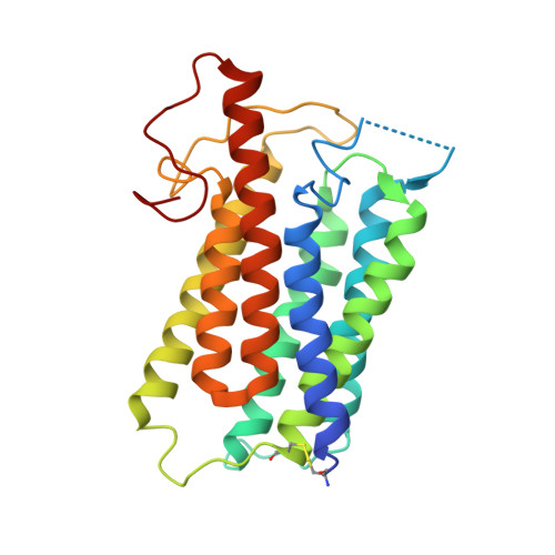
7BW1 | pdb_00007bw1
Crystal structure of Steroid 5-alpha-reductase 2 in complex with Finasteride
- PDB DOI: https://doi.org/10.2210/pdb7BW1/pdb
- Classification: OXIDOREDUCTASE
- Organism(s): Homo sapiens
- Expression System: Baculovirus expression vector pFastBac1-HM
- Mutation(s): No
- Membrane Protein: Yes OPMPDBTMMemProtMD
- Deposited: 2020-04-13 Released: 2020-08-05
- Deposition Author(s): Xiao, Q., Zhang, C., Wei, Z.
- Funding Organization(s): National Natural Science Foundation of China (NSFC)
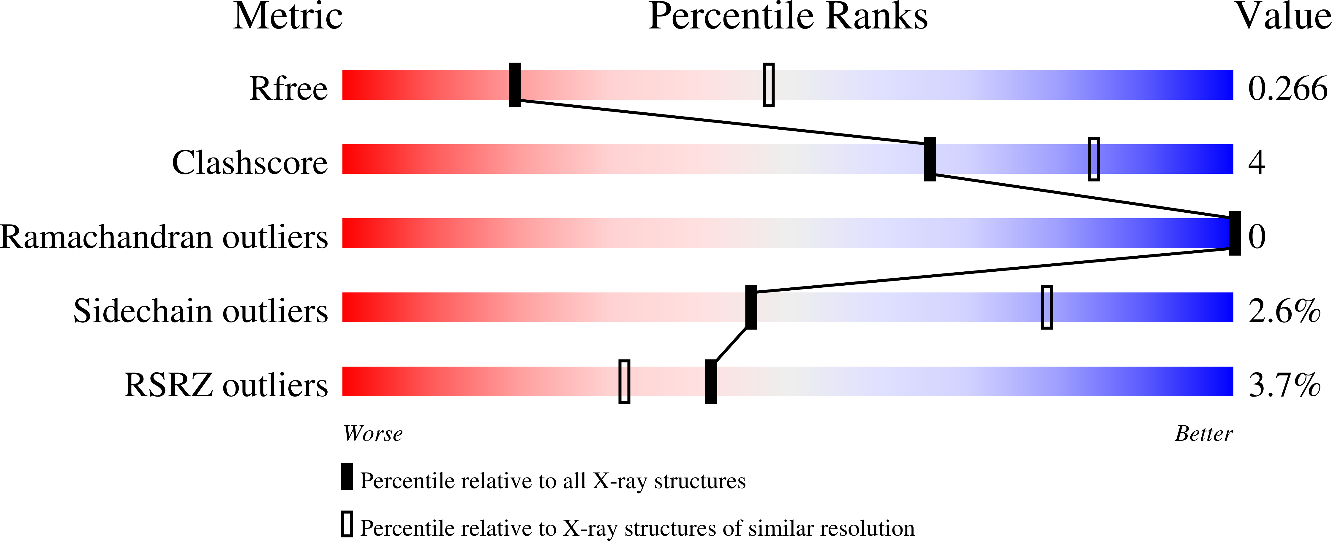
Experimental Data Snapshot
- Method: X-RAY DIFFRACTION
- Resolution: 2.80 Å
- R-Value Free: 0.265 (Depositor), 0.277 (DCC)
- R-Value Work: 0.239 (Depositor), 0.248 (DCC)
- R-Value Observed: 0.241 (Depositor)
Literature
- 👁 Image
Download Mendeley
Structure of human steroid 5 alpha-reductase 2 with anti-androgen drug finasteride.
Xiao, Q., Wang, L., Supekar, S., Shen, T., Liu, H., Ye, F., Huang, J., Fan, H., Wei, Z., Zhang, C.(2020) Res Sq
- PubMed: 32702725 Search on PubMedSearch on PubMed Central
- DOI: https://doi.org/10.21203/rs.3.rs-40159/v1
- Primary Citation Related Structures:
7BW1 - PubMed Abstract:
Human steroid 5α-reductase 2 (SRD5α2) as a critical integral membrane enzyme in steroid metabolism catalyzes testosterone to dihydrotestosterone. Mutations on its gene have been linked to 5α-reductase deficiency and prostate cancer. Finasteride and dutasteride as SRD5α2 inhibitors are widely used anti-androgen drugs for benign prostate hyperplasia, which have recently been indicated in the treatment of COVID-19. The molecular mechanisms underlying enzyme catalysis and inhibition remained elusive for SRD5α2 and other eukaryotic integral membrane steroid reductases due to a lack of structural information. Here, we report a crystal structure of human SRD5α2 at 2.8 Å revealing a unique 7-TM structural topology and an intermediate adduct of finasteride and NADPH as NADP-dihydrofinasteride in a largely enclosed binding cavity inside the membrane. Structural analysis together with computational and mutagenesis studies reveals molecular mechanisms for the 5α-reduction of testosterone and the finasteride inhibition involving residues E57 and Y91. Molecular dynamics simulation results indicate high conformational dynamics of the cytosolic region regulating the NADPH/NADP + exchange. Mapping disease-causing mutations of SRD5α2 to our structure suggests molecular mechanisms for their pathological effects. Our results offer critical structural insights into the function of integral membrane steroid reductases and will facilitate drug development.
- Department of Biology, Southern University of Science and Technology, Shenzhen, Guangdong 518055, China.
- Faculty of Health Sciences, University of Macau, Macau SAR 999078, China.
- Department of Pharmacology and Chemical Biology, School of Medicine, University of Pittsburgh, Pittsburgh, PA15261, USA.
- Bioinformatics Institute (BII), Agency for Science, Technology and Research (ASTAR), Singapore 138671, Singapore.
- Tencent AI Lab, Shenzhen, Guangdong 518000, China.
Organizational Affiliation:
Explore in 3D: Structure | Sequence Annotations | Electron Density | Validation Report | Ligand Interaction (NDX) | Predict Membrane
Biological Assembly 1
Explore in 3D: Structure | Sequence Annotations | Electron Density | Validation Report | Ligand Interaction (NDX) | Predict Membrane
Global Symmetry: Asymmetric - C1
Global Stoichiometry: Monomer - A1
Find Similar Assemblies
Biological assembly 1 assigned by authors and generated by PISA (software)
Biological Assembly Evidence: gel filtration
Macromolecule Content
- Total Structure Weight: 30.36 kDa
- Atom Count: 2,036
- Modeled Residue Count: 245
- Deposited Residue Count: 258
- Unique protein chains: 1
Macromolecules
Entity ID: 1 | |||||
|---|---|---|---|---|---|
| Molecule | Chains | Sequence Length | Organism | Details | Image |
| 3-oxo-5-alpha-steroid 4-dehydrogenase 2 | 258 | Homo sapiens | Mutation(s): 0 Gene Names: SRD5A2 EC: 1.3.1.22 Membrane Entity: Yes | 👁 Image | |
UniProt & NIH Common Fund Data Resources | |||||
PHAROS: P31213 GTEx: ENSG00000277893 | |||||
Entity Groups | |||||
| Sequence Clusters | 30% Identity50% Identity70% Identity90% Identity95% Identity100% Identity | ||||
| UniProt Group | P31213 | ||||
Sequence AnnotationsExpand | |||||
Reference Sequence | |||||
Small Molecules
| Ligands 3 Unique | |||||
|---|---|---|---|---|---|
| ID | Chains | Name / Formula / InChI Key | 2D Diagram | 3D Interactions | |
| NDX (Subject of Investigation/LOI) Download:Ideal Coordinates CCD File | C [auth A] | [[(2~{R},3~{S},4~{R},5~{R})-5-[4-[(1~{S},3~{a}~{S},3~{b}~{S},5~{a}~{R},8~{S},9~{a}~{R},9~{b}~{S},11~{a}~{S})-1-(~{tert}-butylcarbamoyl)-9~{a},11~{a}-dimethyl-7-oxidanylidene-1,2,3,3~{a},3~{b},4,5,5~{a},6,8,9,9~{b},10,11-tetradecahydroindeno[5,4-f]quinolin-8-yl]-3-aminocarbonyl-4~{H}-pyridin-1-yl]-3,4-bis(oxidanyl)oxolan-2-yl]methoxy-oxidanyl-phosphoryl] [(2~{R},3~{R},4~{R},5~{R})-5-(6-aminopurin-9-yl)-3-oxidanyl-4-phosphonooxy-oxolan-2-yl]methyl hydrogen phosphate C44 H66 N9 O19 P3 SBPCUEDRMWGINQ-DAPYLVMHSA-N | 👁 Image | ||
| OLC Download:Ideal Coordinates CCD File | B [auth A] | (2R)-2,3-dihydroxypropyl (9Z)-octadec-9-enoate C21 H40 O4 RZRNAYUHWVFMIP-GDCKJWNLSA-N | 👁 Image | ||
| SO4 Download:Ideal Coordinates CCD File | D [auth A], E [auth A] | SULFATE ION O4 S QAOWNCQODCNURD-UHFFFAOYSA-L | 👁 Image | ||
Experimental Data & Validation
Experimental Data
- Method: X-RAY DIFFRACTION
- Resolution: 2.80 Å
- R-Value Free: 0.265 (Depositor), 0.277 (DCC)
- R-Value Work: 0.239 (Depositor), 0.248 (DCC)
- R-Value Observed: 0.241 (Depositor)
| Length ( Å ) | Angle ( ˚ ) |
|---|---|
| a = 107.449 | α = 90 |
| b = 107.449 | β = 90 |
| c = 103.372 | γ = 120 |
| Software Name | Purpose |
|---|---|
| SCALEPACK | data scaling |
| PHENIX | refinement |
| PDB_EXTRACT | data extraction |
| HKL-2000 | data reduction |
| MOLREP | phasing |
Entry History
& Funding InformationDeposition Data
| Funding Organization | Location | Grant Number |
|---|---|---|
| National Natural Science Foundation of China (NSFC) | China | 31770791 |
| National Natural Science Foundation of China (NSFC) | China | 31971131 |
Revision History (Full details and data files)
- Version 1.0: 2020-08-05
Type: Initial release - Version 1.1: 2024-11-06
Changes: Data collection, Database references, Structure summary