
- FASTA Sequence
- PDBx/mmCIF Format
- PDBx/mmCIF Format (gz)
- BinaryCIF Format (gz)
- Legacy PDB Format
- Legacy PDB Format (gz)
- PDBML/XML Format (gz)
- Structure Factors (CIF)
- Structure Factors (CIF - gz)
- Validation Full (PDF - gz)
- Validation (XML - gz)
- Validation (CIF - gz)
- Validation 2fo-fc coefficients (CIF - gz)
- Validation fo-fc coefficients (CIF - gz)
- Biological Assembly 1 (CIF - gz)
- Biological Assembly 1 (PDB - gz)
7C83 | pdb_00007c83
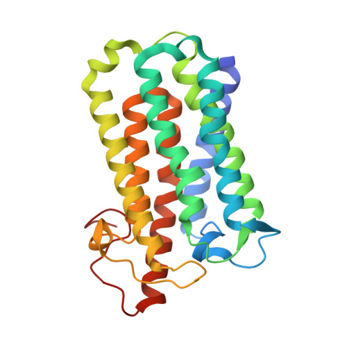
Crystal structure of an integral membrane steroid 5-alpha-reductase PbSRD5A
- PDB DOI: https://doi.org/10.2210/pdb7C83/pdb
- Classification: OXIDOREDUCTASE
- Organism(s): Pseudomonadota bacterium
- Expression System: Spodoptera frugiperda
- Mutation(s): No
- Deposited: 2020-05-28 Released: 2021-01-27
- Deposition Author(s): Ren, R.B., Han, Y.F., Xiao, Q.J., Deng, D.
- Funding Organization(s): National Natural Science Foundation of China (NSFC)
Experimental Data Snapshot
- Method: X-RAY DIFFRACTION
- Resolution: 2.00 Å
- R-Value Free: 0.233 (Depositor), 0.234 (DCC)
- R-Value Work: 0.193 (Depositor), 0.197 (DCC)
- R-Value Observed: 0.197 (Depositor)
Starting Model: in silico
View more details
Literature
- 👁 Image
Download Mendeley
Crystal structure of steroid reductase SRD5A reveals conserved steroid reduction mechanism.
Han, Y., Zhuang, Q., Sun, B., Lv, W., Wang, S., Xiao, Q., Pang, B., Zhou, Y., Wang, F., Chi, P., Wang, Q., Li, Z., Zhu, L., Li, F., Deng, D., Chiang, Y.C., Li, Z., Ren, R.(2021) Nat Commun 12: 449-449
- PubMed: 33469028 Search on PubMedSearch on PubMed Central
- DOI: https://doi.org/10.1038/s41467-020-20675-2
- Primary Citation Related Structures:
7C83 - PubMed Abstract:
Steroid hormones are essential in stress response, immune system regulation, and reproduction in mammals. Steroids with 3-oxo-Δ 4 structure, such as testosterone or progesterone, are catalyzed by steroid 5α-reductases (SRD5As) to generate their corresponding 3-oxo-5α steroids, which are essential for multiple physiological and pathological processes. SRD5A2 is already a target of clinically relevant drugs. However, the detailed mechanism of SRD5A-mediated reduction remains elusive. Here we report the crystal structure of PbSRD5A from Proteobacteria bacterium, a homolog of both SRD5A1 and SRD5A2, in complex with the cofactor NADPH at 2.0 Å resolution. PbSRD5A exists as a monomer comprised of seven transmembrane segments (TMs). The TM1-4 enclose a hydrophobic substrate binding cavity, whereas TM5-7 coordinate cofactor NADPH through extensive hydrogen bonds network. Homology-based structural models of HsSRD5A1 and -2, together with biochemical characterization, define the substrate binding pocket of SRD5As, explain the properties of disease-related mutants and provide an important framework for further understanding of the mechanism of NADPH mediated steroids 3-oxo-Δ 4 reduction. Based on these analyses, the design of therapeutic molecules targeting SRD5As with improved specificity and therapeutic efficacy would be possible.
- Kobilka Institute of Innovative Drug Discovery, School of Life and Health Sciences, The Chinese University of Hong Kong, Shenzhen, Guangdong, 518172, China.
- State Key Laboratory of Cell Biology, Shanghai Institute of Biochemistry and Cell Biology, Center for Excellence in Molecular Cell Science, Chinese Academy of Sciences, University of Chinese Academy of Sciences, 320 Yueyang Road, Shanghai, 200031, China.
- Shanghai Synchrotron Radiation Facility, Shanghai Advanced Research Institute, Chinese Academy of Sciences, Shanghai, 201204, China.
- Warshel Institute for Computational Biology, School of Life and Health Sciences, The Chinese University of Hong Kong, Shenzhen, Guangdong, 518172, China.
- Tencent AI lab, Shenzhen, Guangdong, 518000, China.
- Department of Obstetrics, Key Laboratory of Birth Defects and Related Disease of Women and Children of MOE, State Key Laboratory of Biotherapy, West China Second Hospital, Sichuan University, Chengdu, 610041, China.
- School of Science and Engineering, The Chinese University of Hong Kong, Shenzhen, Guangdong, 518172, China.
- Human Sperm Bank, Key Laboratory of Birth Defects and Related Disease of Women and Children of MOE, West China Second Hospital, Sichuan University, Chengdu, 610041, China.
- Department of Obstetrics, Key Laboratory of Birth Defects and Related Disease of Women and Children of MOE, State Key Laboratory of Biotherapy, West China Second Hospital, Sichuan University, Chengdu, 610041, China. dengd@scu.edu.cn.
- Kobilka Institute of Innovative Drug Discovery, School of Life and Health Sciences, The Chinese University of Hong Kong, Shenzhen, Guangdong, 518172, China. chiangyc@cuhk.edu.cn.
- State Key Laboratory of Cell Biology, Shanghai Institute of Biochemistry and Cell Biology, Center for Excellence in Molecular Cell Science, Chinese Academy of Sciences, University of Chinese Academy of Sciences, 320 Yueyang Road, Shanghai, 200031, China. zhenfei.li@sibcb.ac.cn.
- Kobilka Institute of Innovative Drug Discovery, School of Life and Health Sciences, The Chinese University of Hong Kong, Shenzhen, Guangdong, 518172, China. renruobing@cuhk.edu.cn.
Organizational Affiliation:
Explore in 3D: Structure | Sequence Annotations | Electron Density | Validation Report | Ligand Interaction (NDP)
Biological Assembly 1
Explore in 3D: Structure | Sequence Annotations | Electron Density | Validation Report | Ligand Interaction (NDP)
Global Symmetry: Asymmetric - C1
Global Stoichiometry: Monomer - A1
Find Similar Assemblies
Biological assembly 1 assigned by authors and generated by PISA (software)
Biological Assembly Evidence: gel filtration
Macromolecule Content
- Total Structure Weight: 32.66 kDa
- Atom Count: 2,298
- Modeled Residue Count: 251
- Deposited Residue Count: 258
- Unique protein chains: 1
Macromolecules
Entity ID: 1 | |||||
|---|---|---|---|---|---|
| Molecule | Chains | Sequence Length | Organism | Details | Image |
| 3-oxo-5-alpha-steroid 4-dehydrogenase | 258 | Pseudomonadota bacterium | Mutation(s): 0 Gene Names: DCC71_03205 | 👁 Image | |
Small Molecules
Experimental Data & Validation
Experimental Data
- Method: X-RAY DIFFRACTION
- Resolution: 2.00 Å
- R-Value Free: 0.233 (Depositor), 0.234 (DCC)
- R-Value Work: 0.193 (Depositor), 0.197 (DCC)
- R-Value Observed: 0.197 (Depositor)
| Length ( Å ) | Angle ( ˚ ) |
|---|---|
| a = 52.192 | α = 90 |
| b = 104.266 | β = 90 |
| c = 123.046 | γ = 90 |
| Software Name | Purpose |
|---|---|
| PHENIX | refinement |
| XDS | data reduction |
| Aimless | data scaling |
| PDB_EXTRACT | data extraction |
| PHASER | phasing |
Entry History
& Funding InformationDeposition Data
- Released Date: 2021-01-27 Deposition Author(s): Ren, R.B., Han, Y.F., Xiao, Q.J., Deng, D.
| Funding Organization | Location | Grant Number |
|---|---|---|
| National Natural Science Foundation of China (NSFC) | China | 31971218 |
Revision History (Full details and data files)
- Version 1.0: 2021-01-27
Type: Initial release - Version 1.1: 2021-02-03
Changes: Database references - Version 1.2: 2024-03-27
Changes: Data collection, Database references - Version 1.3: 2024-04-03
Changes: Refinement description