
- FASTA Sequence
- PDBx/mmCIF Format
- PDBx/mmCIF Format (gz)
- BinaryCIF Format (gz)
- Legacy PDB Format
- Legacy PDB Format (gz)
- PDBML/XML Format (gz)
- Structure Factors (CIF)
- Structure Factors (CIF - gz)
- Validation Full (PDF - gz)
- Validation (XML - gz)
- Validation (CIF - gz)
- Validation 2fo-fc coefficients (CIF - gz)
- Validation fo-fc coefficients (CIF - gz)
- Biological Assembly 1 (CIF - gz)
- Biological Assembly 2 (CIF - gz)
- Biological Assembly 1 (PDB - gz)
- Biological Assembly 2 (PDB - gz)
7PCK | pdb_00007pck
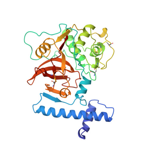
CRYSTAL STRUCTURE OF WILD TYPE HUMAN PROCATHEPSIN K
- PDB DOI: https://doi.org/10.2210/pdb7PCK/pdb
- Classification: HYDROLASE
- Organism(s): Homo sapiens
- Mutation(s): No
- Deposited: 1998-10-21 Released: 1999-10-25
- Deposition Author(s): Sivaraman, J., Lalumiere, M., Menard, R., Cygler, M.
Experimental Data Snapshot
- Method: X-RAY DIFFRACTION
- Resolution: 3.20 Å
- R-Value Free: 0.253 (Depositor), 0.236 (DCC)
- R-Value Work: 0.194 (Depositor), 0.183 (DCC)
- R-Value Observed: 0.194 (Depositor)
Starting Model: experimental
View more details
wwPDB Validation3D Report Full Report
Literature
- 👁 Image
Download Mendeley
Crystal structure of wild-type human procathepsin K.
Sivaraman, J., Lalumiere, M., Menard, R., Cygler, M.(1999) Protein Sci 8: 283-290
- PubMed: 10048321 Search on PubMedSearch on PubMed Central
- DOI: https://doi.org/10.1110/ps.8.2.283
- Primary Citation Related Structures:
7PCK - PubMed Abstract:
Cathepsin K is a lysosomal cysteine protease belonging to the papain superfamily. It has been implicated as a major mediator of osteoclastic bone resorption. Wild-type human procathepsin K has been crystallized in a glycosylated and a deglycosylated form. The latter crystals diffract better, to 3.2 A resolution, and contain four molecules in the asymmetric unit. The structure was solved by molecular replacement and refined to an R-factor of 0.194. The N-terminal fragment of the proregion forms a globular domain while the C-terminal segment is extended and shows substantial flexibility. The proregion interacts with the enzyme along the substrate binding groove and along the proregion binding loop (residues Ser138-Asn156). It binds to the active site in the opposite direction to that of natural substrates. The overall binding mode of the proregion to cathepsin K is similar to that observed in cathepsin L, caricain, and cathepsin B, but there are local differences that likely contribute to the specificity of these proregions for their cognate enzymes. The main observed difference is in the position of the short helix alpha3p (67p-75p), which occupies the S' subsites. As in the other proenzymes, the proregion utilizes the S2 subsite for anchoring by placing a leucine side chain there, according to the specificity of cathepsin K toward its substrate.
- Biotechnology Research Institute, National Research Council of Canada, Montréal, Québec.
Organizational Affiliation:
Explore in 3D: Structure | Sequence Annotations | Electron Density | Validation Report
Biological Assembly 1
Explore in 3D: Structure | Sequence Annotations | Electron Density | Validation Report
Global Symmetry: Cyclic - C2 (Explore in 3D)
Global Stoichiometry: Homo 2-mer - A2
Find Similar Assemblies
Biological assembly 1 assigned by authors and generated by PISA (software)
Biological Assembly 2
Explore in 3D: Structure | Sequence Annotations | Electron Density | Validation Report
Global Symmetry: Cyclic - C2 (Explore in 3D)
Global Stoichiometry: Homo 2-mer - A2
Find Similar Assemblies
Biological assembly 2 assigned by authors and generated by PISA (software)
Macromolecule Content
- Total Structure Weight: 141.43 kDa
- Atom Count: 9,305
- Modeled Residue Count: 1,232
- Deposited Residue Count: 1,256
- Unique protein chains: 1
Macromolecules
Entity ID: 1 | |||||
|---|---|---|---|---|---|
| Molecule | Chains | Sequence Length | Organism | Details | Image |
| PROTEIN (PROCATHEPSIN K) | 314 | Homo sapiens | Mutation(s): 0 EC: 3.4.22.38 | 👁 Image | |
UniProt & NIH Common Fund Data Resources | |||||
PHAROS: P43235 GTEx: ENSG00000143387 | |||||
Entity Groups | |||||
| Sequence Clusters | 30% Identity50% Identity70% Identity90% Identity95% Identity100% Identity | ||||
| UniProt Group | P43235 | ||||
Sequence AnnotationsExpand | |||||
Reference Sequence | |||||
Experimental Data & Validation
Experimental Data
- Method: X-RAY DIFFRACTION
- Resolution: 3.20 Å
- R-Value Free: 0.253 (Depositor), 0.236 (DCC)
- R-Value Work: 0.194 (Depositor), 0.183 (DCC)
- R-Value Observed: 0.194 (Depositor)
| Length ( Å ) | Angle ( ˚ ) |
|---|---|
| a = 58.7 | α = 90 |
| b = 84.4 | β = 90.3 |
| c = 155.6 | γ = 90 |
| Software Name | Purpose |
|---|---|
| DENZO | data reduction |
| SCALEPACK | data scaling |
| AMoRE | phasing |
| X-PLOR | refinement |
Entry History
Deposition Data
- Released Date: 1999-10-25 Deposition Author(s): Sivaraman, J., Lalumiere, M., Menard, R., Cygler, M.
Revision History (Full details and data files)
- Version 1.0: 1999-10-25
Type: Initial release - Version 1.1: 2008-04-27
Changes: Version format compliance - Version 1.2: 2011-07-13
Changes: Version format compliance - Version 1.3: 2020-01-29
Changes: Derived calculations - Version 1.4: 2023-09-20
Changes: Data collection, Database references, Refinement description - Version 1.5: 2024-11-20
Changes: Structure summary