
- FASTA Sequence
- PDBx/mmCIF Format
- PDBx/mmCIF Format (gz)
- BinaryCIF Format (gz)
- Legacy PDB Format
- Legacy PDB Format (gz)
- PDBML/XML Format (gz)
- Structure Factors (CIF)
- Structure Factors (CIF - gz)
- Validation Full (PDF - gz)
- Validation (XML - gz)
- Validation (CIF - gz)
- Validation 2fo-fc coefficients (CIF - gz)
- Validation fo-fc coefficients (CIF - gz)
- Biological Assembly 1 (CIF - gz)
- Biological Assembly 1 (PDB - gz)
3FPU | pdb_00003fpu
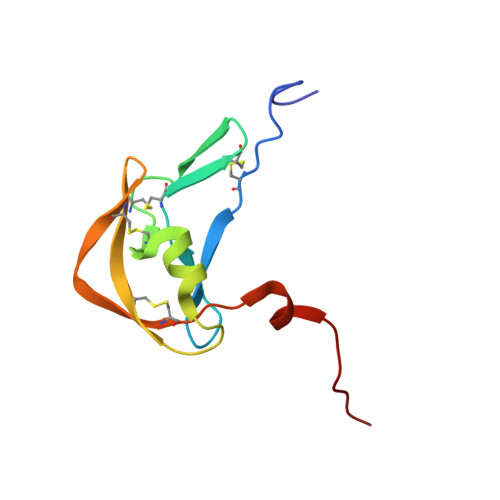
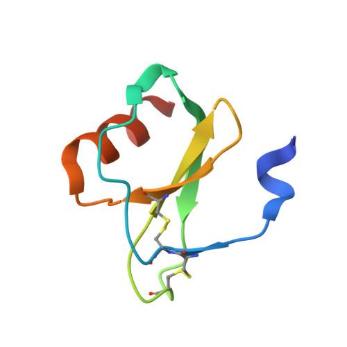
The crystallographic structure of the Complex between Evasin-1 and CCL3
- PDB DOI: https://doi.org/10.2210/pdb3FPU/pdb
- Classification: IMMUNE SYSTEM
- Organism(s): Rhipicephalus sanguineus, Homo sapiens
- Expression System: Spodoptera frugiperda, Escherichia coli
- Mutation(s): Yes
- Deposited: 2009-01-06 Released: 2010-01-12
- Deposition Author(s): Shaw, J.P., Dias, J.M.
Experimental Data Snapshot
- Method: X-RAY DIFFRACTION
- Resolution: 1.76 Å
- R-Value Free: 0.285 (Depositor), 0.280 (DCC)
- R-Value Work: 0.231 (Depositor), 0.230 (DCC)
- R-Value Observed: 0.234 (Depositor)
Starting Model: experimental
View more details
wwPDB Validation 3D Report Full Report
- 👁 Image
Download Mendeley
Structural basis of chemokine sequestration by a tick chemokine binding protein: the crystal structure of the complex between Evasin-1 and CCL3
Dias, J.M., Losberger, C., Deruaz, M., Power, C.A., Proudfoot, A.E.I., Shaw, J.P.(2009) PLoS One 4
- PubMed: 20041127 Search on PubMedSearch on PubMed Central
- DOI: https://doi.org/10.1371/journal.pone.0008514
- Primary Citation Related Structures:
3FPR, 3FPT, 3FPU - PubMed Abstract:
Chemokines are a subset of cytokines responsible for controlling the cellular migration of inflammatory cells through interaction with seven transmembrane G protein-coupled receptors. The blocking of a chemokine-receptor interaction results in a reduced inflammatory response, and represents a possible anti-inflammatory strategy, a strategy that is already employed by some virus and parasites. Anti-chemokine activity has been described in the extracts of tick salivary glands, and we have recently described the cloning and characterization of such chemokine binding proteins from the salivary glands, which we have named Evasins. We have solved the structure of Evasin-1, a very small and highly selective chemokine-binding protein, by x-ray crystallography and report that the structure is novel, with no obvious similarity to the previously described structures of viral chemokine binding proteins. Moreover it does not possess a known fold. We have also solved the structure of the complex of Evasin-1 and its high affinity ligand, CCL3. The complex is a 1:1 heterodimer in which the N-terminal region of CCL3 forms numerous contacts with Evasin-1, including prominent pi-pi interactions between residues Trp89 and Phe14 of the binding protein and Phe29 and Phe13 of the chemokine. However, these interactions do not appear to be crucial for the selectivity of the binding protein, since these residues are found in CCL5, which is not a ligand for Evasin-1. The selectivity of the interaction would appear to lie in the N-terminal residues of the chemokine, which form the "address" whereas the hydrophobic interactions in the rest of the complex would serve primarily to stabilize the complex. A thorough understanding of the binding mode of this small protein, and its other family members, could be very informative in the design of potent neutralizing molecules of pro-inflammatory mediators of the immune system, such as chemokines.
- Merck Serono Geneva Research Center, Merck Serono S.A., Geneva, Switzerland.
Organizational Affiliation:
Explore in 3D: Structure | Sequence Annotations | Electron Density | Validation Report | Ligand Interaction (NI)
Explore in 3D: Structure | Sequence Annotations | Electron Density | Validation Report | Ligand Interaction (NI)
Global Symmetry: Asymmetric - C1
Global Stoichiometry: Hetero 2-mer - A1B1
Find Similar Assemblies
Biological assembly 1 assigned by authors and generated by PISA (software)
Macromolecule Content
- Total Structure Weight: 19.31 kDa
- Atom Count: 1,470
- Modeled Residue Count: 166
- Deposited Residue Count: 170
- Unique protein chains: 2
Entity ID: 1 | |||||
|---|---|---|---|---|---|
| Molecule | Chains | Sequence Length | Organism | Details | Image |
| Evasin-1 | 100 | Rhipicephalus sanguineus | Mutation(s): 0 | 👁 Image | |
UniProt | |||||
Find proteins for P0C8E7 (Rhipicephalus sanguineus) Explore P0C8E7 Go to UniProtKB: P0C8E7 | |||||
Entity Groups | |||||
| Sequence Clusters | 30% Identity50% Identity70% Identity90% Identity95% Identity100% Identity | ||||
| UniProt Group | P0C8E7 | ||||
Sequence AnnotationsExpand | |||||
| |||||
Entity ID: 2 | |||||
|---|---|---|---|---|---|
| Molecule | Chains | Sequence Length | Organism | Details | Image |
| C-C motif chemokine 3 | 70 | Homo sapiens | Mutation(s): 1 | 👁 Image | |
UniProt & NIH Common Fund Data Resources | |||||
Find proteins for P10147 (Homo sapiens) Explore P10147 Go to UniProtKB: P10147 | |||||
PHAROS: P10147 GTEx: ENSG00000277632 | |||||
Entity Groups | |||||
| Sequence Clusters | 30% Identity50% Identity70% Identity90% Identity95% Identity100% Identity | ||||
| UniProt Group | P10147 | ||||
Sequence AnnotationsExpand | |||||
| |||||
| Ligands 1 Unique | |||||
|---|---|---|---|---|---|
| ID | Chains | Name / Formula / InChI Key | 2D Diagram | 3D Interactions | |
| NI Query on NI Download Ideal Coordinates CCD File | C [auth A], D [auth A] | NICKEL (II) ION Ni VEQPNABPJHWNSG-UHFFFAOYSA-N | 👁 Image | ||
Experimental Data
- Method: X-RAY DIFFRACTION
- Resolution: 1.76 Å
- R-Value Free: 0.285 (Depositor), 0.280 (DCC)
- R-Value Work: 0.231 (Depositor), 0.230 (DCC)
- R-Value Observed: 0.234 (Depositor)
| Length ( Å ) | Angle ( ˚ ) |
|---|---|
| a = 104.384 | α = 90 |
| b = 104.384 | β = 90 |
| c = 104.384 | γ = 90 |
| Software Name | Purpose |
|---|---|
| HKL-2000 | data collection |
| AMoRE | phasing |
| REFMAC | refinement |
| DENZO | data reduction |
| SCALEPACK | data scaling |
Deposition Data
- Released Date: 2010-01-12 Deposition Author(s): Shaw, J.P., Dias, J.M.
Revision History (Full details and data files)
- Version 1.0: 2010-01-12
Type: Initial release - Version 1.1: 2011-07-13
Changes: Version format compliance - Version 1.2: 2013-10-16
Changes: Derived calculations - Version 1.3: 2021-11-10
Changes: Database references, Derived calculations - Version 1.4: 2023-11-01
Changes: Data collection, Refinement description - Version 1.5: 2024-11-20
Changes: Structure summary