
- FASTA Sequence
- PDBx/mmCIF Format
- PDBx/mmCIF Format (gz)
- BinaryCIF Format (gz)
- Legacy PDB Format
- Legacy PDB Format (gz)
- PDBML/XML Format (gz)
- Structure Factors (CIF)
- Structure Factors (CIF - gz)
- Validation Full (PDF - gz)
- Validation (XML - gz)
- Validation (CIF - gz)
- Validation 2fo-fc coefficients (CIF - gz)
- Validation fo-fc coefficients (CIF - gz)
- Biological Assembly 1 (CIF - gz)
- Biological Assembly 2 (CIF - gz)
- Biological Assembly 3 (CIF - gz)
- Biological Assembly 1 (PDB - gz)
- Biological Assembly 2 (PDB - gz)
- Biological Assembly 3 (PDB - gz)
4RA8 | pdb_00004ra8
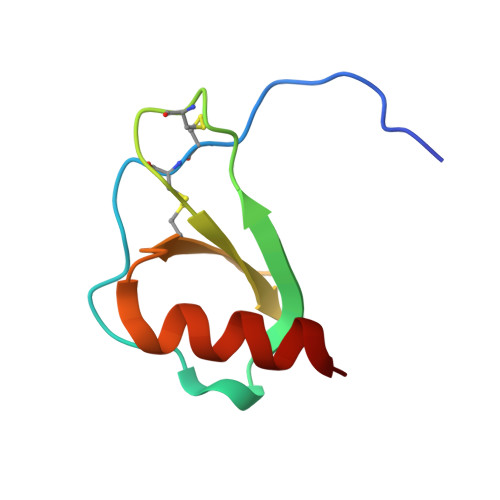
Structure analysis of the Mip1a P8A mutant
- PDB DOI: https://doi.org/10.2210/pdb4RA8/pdb
- Entry: 4RA8 supersedes: 3TN1
- Classification: CYTOKINE
- Organism(s): Homo sapiens
- Expression System: Escherichia coli
- Mutation(s): Yes
- Deposited: 2014-09-09 Released: 2014-09-24
- Deposition Author(s): Liang, W.G., Ren, M., Guo, Q., Tang, W.J.
Experimental Data Snapshot
- Method: X-RAY DIFFRACTION
- Resolution: 2.60 Å
- R-Value Free: 0.248 (Depositor), 0.240 (DCC)
- R-Value Work: 0.202 (Depositor), 0.210 (DCC)
- R-Value Observed: 0.204 (Depositor)
Starting Model: experimental
View more details
wwPDB Validation 3D Report Full Report
- 👁 Image
Download Mendeley
Structures of human CCL18, CCL3, and CCL4 reveal molecular determinants for quaternary structures and sensitivity to insulin-degrading enzyme.
Liang, W.G., Ren, M., Zhao, F., Tang, W.J.(2015) J Mol Biology 427: 1345-1358
- PubMed: 25636406 Search on PubMedSearch on PubMed Central
- DOI: https://doi.org/10.1016/j.jmb.2015.01.012
- Primary Citation Related Structures:
3TN2, 4MHE, 4RA8, 4RAL - PubMed Abstract:
CC chemokine ligands (CCLs) are 8- to 14-kDa signaling proteins involved in diverse immune functions. While CCLs share similar tertiary structures, oligomerization produces highly diverse quaternary structures that protect chemokines from proteolytic degradation and modulate their functions. CCL18 is closely related to CCL3 and CCL4 with respect to both protein sequence and genomic location, yet CCL18 has distinct biochemical and biophysical properties. Here, we report a crystal structure of human CCL18 and its oligomerization states in solution based on crystallographic and small-angle X-ray scattering analyses. Our data show that CCL18 adopts an α-helical conformation at its N-terminus that weakens its dimerization, explaining CCL18's preference for the monomeric state. Multiple contacts between monomers allow CCL18 to reversibly form a unique open-ended oligomer different from those of CCL3, CCL4, and CCL5. Furthermore, these differences hinge on proline 8, which is conserved in CCL3 and CCL4 but is replaced by lysine in human CCL18. Our structural analyses suggest that a mutation of proline 8 to alanine stabilizes a type 1 β-turn at the N-terminus of CCL4 to prevent dimerization but prevents dimers from making key contacts with each other in CCL3. Thus, the P8A mutation induces depolymerization of CCL3 and CCL4 by distinct mechanisms. Finally, we used structural, biochemical, and functional analyses to unravel why insulin-degrading enzyme degrades CCL3 and CCL4 but not CCL18. Our results elucidate the molecular basis for the oligomerization of three closely related CC chemokines and suggest how oligomerization shapes CCL chemokine function.
- Ben May Department for Cancer Research, The University of Chicago, IL 60637, USA.
- Ben May Department for Cancer Research, The University of Chicago, IL 60637, USA. Electronic address: wtang@uchicago.edu.
Organizational Affiliation:
Explore in 3D: Structure | Sequence Annotations | Electron Density | Validation Report
Explore in 3D: Structure | Sequence Annotations | Electron Density | Validation Report
Global Symmetry: Cyclic - C2 (Explore in 3D)
Global Stoichiometry: Homo 2-mer - A2
Find Similar Assemblies
Biological assembly 1 assigned by authors and generated by PISA (software)
Explore in 3D: Structure | Sequence Annotations | Electron Density | Validation Report
Global Symmetry: Cyclic - C2 (Explore in 3D)
Global Stoichiometry: Homo 2-mer - A2
Find Similar Assemblies
Biological assembly 2 assigned by authors and generated by PISA (software)
Explore in 3D: Structure | Sequence Annotations | Electron Density | Validation Report
Global Symmetry: Cyclic - C2 (Explore in 3D)
Global Stoichiometry: Homo 2-mer - A2
Find Similar Assemblies
Biological assembly 3 assigned by authors and generated by PISA (software)
Macromolecule Content
- Total Structure Weight: 38.48 kDa
- Atom Count: 2,691
- Modeled Residue Count: 336
- Deposited Residue Count: 345
- Unique protein chains: 1
Entity ID: 1 | |||||
|---|---|---|---|---|---|
| Molecule | Chains | Sequence Length | Organism | Details | Image |
| C-C motif chemokine 3 | 69 | Homo sapiens | Mutation(s): 1 Gene Names: CCL3, G0S19-1, MIP1A, SCYA3 | 👁 Image | |
UniProt & NIH Common Fund Data Resources | |||||
Find proteins for P10147 (Homo sapiens) Explore P10147 Go to UniProtKB: P10147 | |||||
PHAROS: P10147 GTEx: ENSG00000277632 | |||||
Entity Groups | |||||
| Sequence Clusters | 30% Identity50% Identity70% Identity90% Identity95% Identity100% Identity | ||||
| UniProt Group | P10147 | ||||
Sequence AnnotationsExpand | |||||
| |||||
Experimental Data
- Method: X-RAY DIFFRACTION
- Resolution: 2.60 Å
- R-Value Free: 0.248 (Depositor), 0.240 (DCC)
- R-Value Work: 0.202 (Depositor), 0.210 (DCC)
- R-Value Observed: 0.204 (Depositor)
| Length ( Å ) | Angle ( ˚ ) |
|---|---|
| a = 180.154 | α = 90 |
| b = 180.154 | β = 90 |
| c = 77.548 | γ = 120 |
| Software Name | Purpose |
|---|---|
| HKL-3000 | data collection |
| PHENIX | model building |
| PHENIX | refinement |
| HKL-3000 | data reduction |
| HKL-3000 | data scaling |
| PHENIX | phasing |
Deposition Data
- Released Date: 2014-09-24 Deposition Author(s): Liang, W.G., Ren, M., Guo, Q., Tang, W.J.
- This entry supersedes: 3TN1
Revision History (Full details and data files)
- Version 1.0: 2014-09-24
Type: Initial release - Version 1.1: 2015-05-13
Changes: Database references - Version 1.2: 2017-11-22
Changes: Refinement description - Version 1.3: 2023-09-20
Changes: Data collection, Database references, Refinement description - Version 1.4: 2024-11-27
Changes: Structure summary